Lungs
- Lungs are essential organ of respiration.
- Shape of lungs are like half-cone.
- Lungs has an Apex & Base and 2 Surfaces and 3 Borders.
- Two lungs are present in every human body.
 |
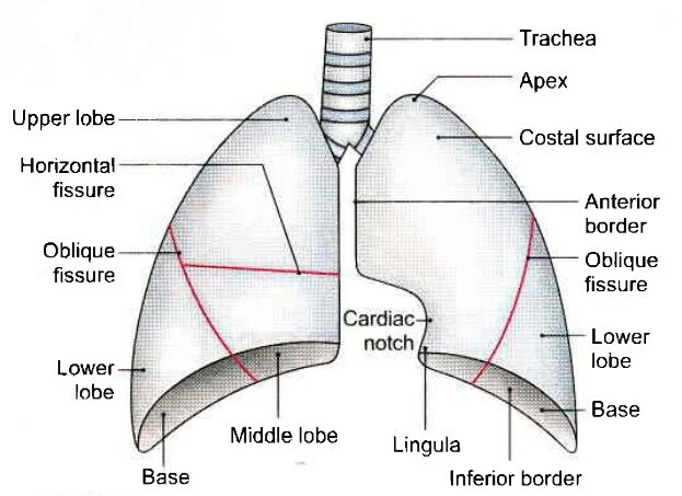
| The trachea and lungs as seen from the front |
Base -
- It is deeply concave and faces downwards.
- It rest over the dom of diaphragm.
Apex -
- It is conical & directed upwards, towards the root of the neck.
Borders (3) -
Anterior Border
- It is sharp and related to the anterior thoracic wall.
- It's lower part has deep concavity of the left side which is called as cardiac notch.
Posterior Border
- It is rounded & found the junction of costal and medial surface.
- It corresponds with the head of the ribs.
Inferior Border
- It is convex and sharp, it faces downwards.
Surfaces (2) -
Costal Surface
- This surface is related to rib cage & sternum.
Medial Surface
It is divided into two parts...
1. Vertibral part - it is the posterior part of the medial surface and related to the vertibral bodies.
2. Mediastnal part - this surface is marked by hilum ( Hilum - it is depression from which structure enters or exit from here).
It is the large anterior part of the medial surface which is related to the mediastinum.
Side Determination -
- Sharp anterior border faces forward.
- Deep concave faces inferiorly.
- Surface having hilum faces medially.
Right Lung -
- It has 2 fissures (Oblique, Transverse).
- It has 3 lobes ( Superior, Middle and Inferior).
 |
| Impression on the mediastinal surface of right lung |
Left Lung -
- It has only one fissure ( Oblique).
- It has 2 lobes (Superior and Inferior).
- The lower part of superior lobe is also called as lingula, which correspond with middle lobe of the right lung.
Impression on the mediastinal surface of left lung |
Difference between Right and Left Lung...
Right Lung Left Lung
1. Weight- 625gm - 565gm
2. Size - Shorter & broader- longer & narrower
3. Fissure- Two - one
4. Lobes - Three - Two
5. Lingula - absent - present
6. Cardiac notch - absent - present
7. BPS - 10 - 8
Broncho pulmonary segments (BPS)
- It is difine as the developmental structural and functional unit of the lungs.
- It consists of tersory bronchus and lungs tissue.
Right lung (10 - BPS)
- Superior lobe has 3 BPS
- Middle lobe has 2 BPS
- Inferior lobe has 5 BPS
Left lung ( 8 - BPS)
- Superior lobe has 3 BPS
- Inferior lobe has 5 BPS
 |
| Bronchiopulmonary segments |
Arterial Supply of Lungs
1. Pulmonary Arteries - one on each side ( Right and Left lungs).
2. Bronchial Arteries - one for right lung and two for left lung.
Venous drainage
1. Pulmonary vein - two from right and two from left lungs (a superior and a inferior).
2. Bronchial vein - one from each lung.
Lymphatic drainage
1. Pulmonary limph node.
2. Hilar limph node.
3. Trachiobronchial limph node.
4. Paratrachial limph node.
Nerve Supply
- Sympathetic - anterior and posterior pulmonary plexus.
- Parasympathetic - vagus nerve.
Applied
1. Pneumonectomy - removal of whole lung.
2. Lobectomy - removal of lobe.
3. Segmental resection - removal of BPS.


Thanks for your feedback
ReplyDelete