Showing posts with label General. Show all posts

Overview of neoplasm

Definition

A mass of tissue formed as a result of abnormal, excessive, uncoordinated, autonomous, and purposeless proliferation of cells even after removal of stimulus. 


  • Neoplasm is a term while neoplasia is a process. 
  • Tumour have two components, one is parenchyma (neoplastic cell) and second is stroma (supporting cells and vessels).

Nucleus

Name :- Nucleus (Nucleus was first described by Robert Brown in 1831, and it's material stained by the basic dyes was given the name chromatin by Flemming.)


Properties :

  • It is the largest cellular organelle.
  • Generally located in the center of the cell but it may vary in some cells.
  • It's diameter is about 10µ to 22µ.
  • Mostly it is spherical in shape but it may vary in some cells.
  • Nucleus occupies about 10% of total volume of the cell.
  • It is present in all the cells in the body except RBCs.
  • The cells with nucleus called eukaryotes and without nucleus called prokaryotes.
  • Presence of nucleus is necessary for cell division.
  • Most of the cells have only one nucleus called uninucleated cells.
  • Few types of cells like skeletal muscle have many nuclei called multinucleated cells.
Nucleus


Structure Of Nucleus :
  1. Nuclear membrane
  2. Major components like nucleoplasm, chromatin, chromosomes and nucleolus.
 1. Nuclear Membrane (NM) :

  • NM is double layered membrane called nuclear envelope, separates the nucleus from the cytoplasm.
  • NM is porous in nature, which allows the nucleoplasm to communicate with the cytoplasm.
  • Pores (diameter - 80-100 nm) of the NM are lined by protein molecules (after attachment of protein molecules, pores diameter may decrease 7-9 nm).
  • Small molecules and ions move through the pores by passive diffusion, while large molecules such as RNAs and proteins pass through the selectively active transport.
  • Outer membrane of NM is continue with membrane of endoplasmic reticulum (ER).
  • The space between two layered of NM is continue with the lumen of the ER.
2. Major Components : Such as nucleoplasm, chromatin, chromosomes and nucleolus ....

Nucleoplasm :
  • Nucleoplasm is a highly viscous fluid similar to cytoplasm.
  • It surrounds chromatin and nucleolus.
  • It contains nuclear matrix (dense fibrillar network of protein) and many substances such as nucleotides, enzymes and hyaloplasm (soluble liquid part of nucleoplasm).
  • Nuclear matrix forms the structural framework for organizing chromatin.
Chromatin : 
  • Chromatin is a thread like material referred as DNA-histone complex or complex of DNA, proteins (histones) and some RNA. (A chain of nucleosomes)
  • DNA is a double helix which wraps around the central core of eight histone molecules to form the fundamental packing unit of chromatin called nucleosome. (Eight histones wrapped by double helix DNA)
  • Nucleosomes are packed together tightly with the help of histone molecule to form a chromatin fiber.
  • Just before cell division, the chromatin condenses to form a pair of chromatids that constitutes a chromosome.
  • In the cell that are not dividing, the chromatin appears as a diffuse granular mass.

Nucleus


Chromosomes : 
  • Chromosome is the rod-shaped nuclear structure that carries a complete blueprint of all the hereditary characteristics of that species.
  • Each chromosome is a long molecule of DNA (contains many genes) that is coiled together with several proteins.
  • Normally, the chromosomes are not visible in the nucleus under microscope while it visible (because DNA more tightly packed just before cell division) only during cell division.
  • All the dividing cells of the body except reproductive cells contain 23 pairs of chromosomes.
  • Each pair consists of one chromosome inherited from father and one from mother.
  • The cells with 23 pairs of chromosomes are called diploid cells. The cells with 23 single chromosomes called haploid cells like reproductive cells also called for this gametes or sex cells
Nucleolus (pleural - nucleoli) :
  • Nucleolus is a small, round granular structure of the nucleus.
  • Each nucleus contain one or more nucleoli.
  • Nucleoli are quite prominent in cells that synthesize large amount of protein like muscle and liver cells.
  • Nucleoli disperse and disappear during cell division and reorganize once new cells are formed.
  • The nucleolus contains RNA and some proteins which are similar to those found in ribosomes.
  • The RNA is synthesized by five different pairs of chromosomes and stored in nucleolus. Later, it condensed to form sub-units of ribosomes. All the sub-units are transported from nucleolus to cytoplasm through pores of the nuclear membrane.
Functions Of Nucleus :
  1. Control of the all activities of the cell such as metabolism, protein synthesis, growth and reproduction (cell division).
  2. Synthesis of RNA.
  3. Formation of sub-units of ribosomes.
  4. Sending genetic instruction to the cytoplasm for protein synthesis through messenger RNA (mRNA).
  5. Control of the cell division through genes.
  6. Storage of hereditary information (in genes) and transformation of this information from one generation of the species to the next.

Similar Posts :

Golgi apparatus

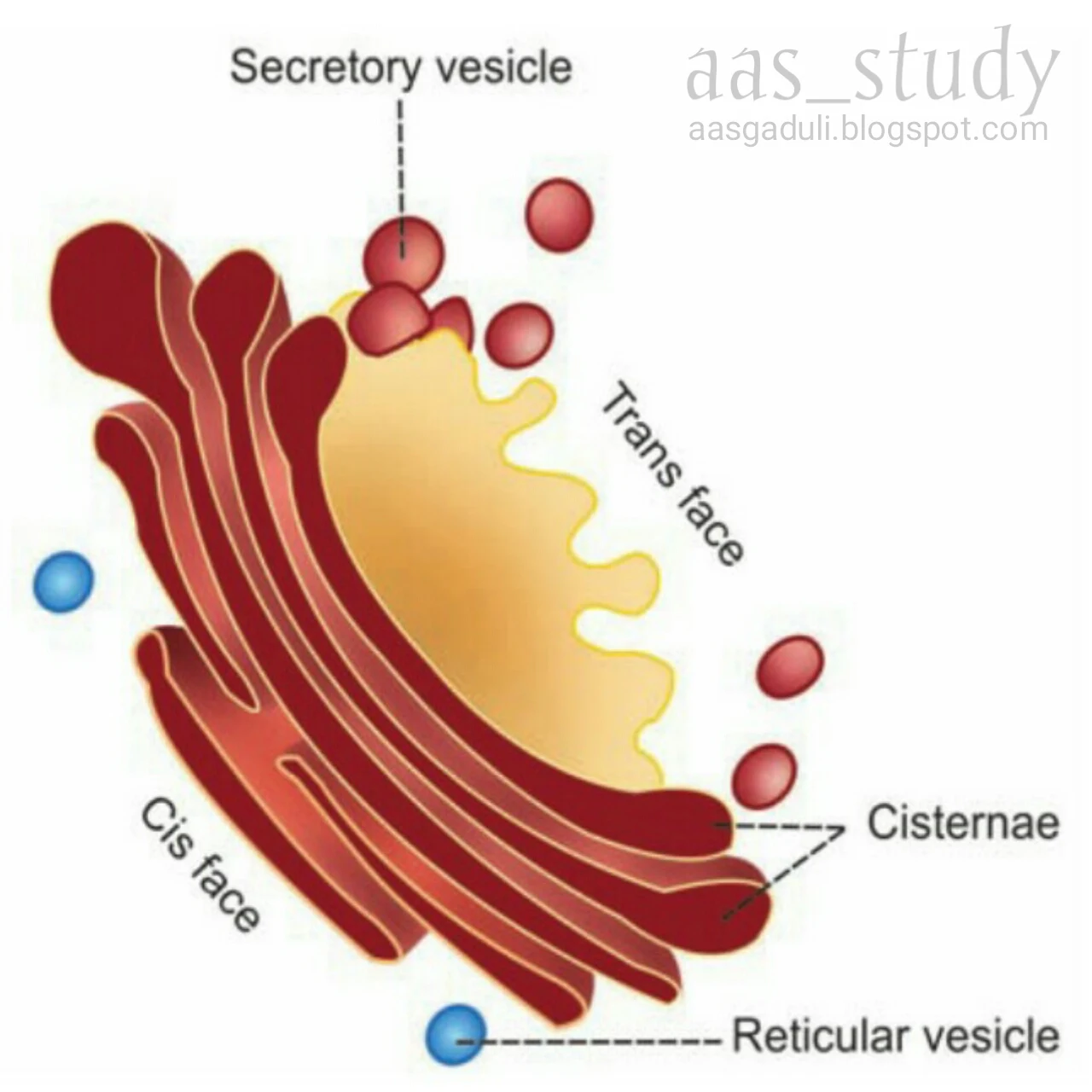
Name :- Golgi apparatus, Golgi body, Golgi complex, Post office of the cell, Shipping department of the cell (Camillo Golgi (1898) first discovered, hence the name is Golgi).

Properties :
  • It is an membrane bound organelle situated near the nucleus.
  • It is present in all cells except RBCs.
  • Usually, each cell has one Golgi apparatus.
  • Each Golgi apparatus consists of 5-8 flattened membranous sacs called cisternae (0.5-1.0 micrometer).
  • Golgi apparatus has two ends or faces, namely cis face (situated near the endoplasmic reticulum) and trans face (situated near the cell membrane).
Functions :
Major functions of Golgi apparatus are processing, packing, labeling and delivery of proteins and lipids to different part of the cell or outside of the cell.
  1. Processing :- glycoproteins and lipids are modified here.
  2. Packaging :- modified materials are packed in the form of secretory granules, secretory vesicles and lysosomes.
  3. Labeling and delivery :- Golgi apparatus sort out the processed materials and labels them (such as phosphate group) then distribute in or out side of the cell according to demand.

Golgi apparatus


Similar Posts :-

Cell Adaptation

Adaptive Disorders

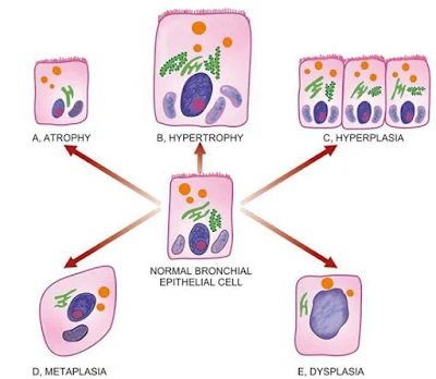
Definition :- Cell become change or adapt in response to adverse environmental changes that's called adaptation.

Types of adaptation
1. Atrophy (decrease cell size)
2. Dysplasia ( abnormal growth or development of cell )
3. Hyperplasia ( increase reproduction rate of the cell )
4. Hypertrophy ( increase cell size )
5. Metaplasia ( conversion of the cell from one type to another type e.g. squamus cell convert into columnar cell )

Adaptive Disorders

Difference between Metaplasia and Dysplasia


Similar Posts -


Dysplasia / Atypical hyperplasia

Definition – It is the disordered cellular development associated with metaplasia and hyperplasia caused by chronic irritation and prolonged inflammation. It is occur most commonly in epithelial cells.

Dysplasia characterised by cellular proliferation and cytologic changes as under…
  • Increase number of layers of epithelial cells.
  • Disorderly arrangement of cells from basal layer to the surface layer.
  • Loss of polarity i.e. nuclei lying away from basement membrane.
  • Cellular and nuclear pleomorphism.
  • Increase nucleocytoplasmic ratio.
  • Nuclear hyperchromatism.
  • Increase mitotic activity.

Most common examples of dysplastic changes are the uterine cervix and respiratory tract.

Metaplasia, dysplasia, adaptation , aasgaduli, pathology
Difference between Metaplasia and Dysplasia


Similar Posts -

Metaplasia


Definition – Metaplasia is a reversible condition in which one type of epithelial or mesenchymal adult cells convert into another type of epithelial or mesenchymal cells, usually in response to abnormal stimuli and it reverts back after removal of stimulus. If stimulus persists for long time then metaplasia convert into dysplasia and further convert into cancer.
Types – (1) Epithelial metaplasia (2) Mesenchymal metaplasia
1. Epithelial metaplasia – This is the more common type in which metaplastic changes may be patchy or diffuse and usually replaced by stronger but less well-specialised epithelium. There are two types of epithelial metaplasia…
A. SQUAMOUS METAPLASIA – It is caused by chronic irritation that may be mechanical, chemical or infective in origin. Some common examples…
  • In bronchus (normally lined by pseudostratified columnar ciliated epithelium) in chronic smokers.
  • In uterine endocervix (normally lined by simple columnar epithelium) in prolapsed of the uterus and in old age.
  • In gallbladder (normally lined by simple columnar epithelium) in chronic cholecystitis and cholelithiasis.
  • In prostate (ducts normally lined by simple columnar epithelium) in chronic prostatitis and oestrogen therapy.
  • In renal pelvis and urinary bladder (normally lined by transitional epithelium) in chronic infection and stones.
  • In vitamin A deficiency (xerophthalmia) in nose, bronchi, urinary tract, lacrimal and salivary glands.

B. COLUMNAR METAPLASIA – There are some conditions in which there is transformation to columnar epithelium. For example_
  • Intestinal metaplasia in healed chronic gastric ulcer.
  • Columnar metaplasia in Barrett’s oesophagus (change squamous to columnar epithelium).
  • Pseudostratified ciliated columnar epithelium change into columnar epithelium in chronic bronchitis and bronchiectasis.

2. Mesenchymal metaplasia – Less often, there is transformation of one adult type of mesenchymal tissue to another. There is two types
A. OSSEOUS METAPLASIA – Formation of bone in fibrous tissue, cartilage and myxoid tissue. Examples…
  • In arterial wall in old age (Monckeberg’s medial calcific sclerosis).
  • In soft tissues in myositis ossificans.
  • In cartilage of larynx and bronchi in elderly people.
  • In scar of chronic inflammation of prolonged duration.

B. CARTILAGINOUS METAPLASIA – It may occur in healing of fractures, where there is undue mobility.

Metaplasia, dysplasia, adaptation, pathology
Differences between Metaplasia and Dysplasia


Similar Posts -

Hyperplasia


Definition – Hyperplasia is an increase in the number of parenchymal cells resulting in enlargement of the organ or tissue. Quite often, both hyperplasia and hypertrophy occur together.
Causes – It may be physiological or pathological…
PHYSIOLOGICAL HYPERPLASIA –
                  1. Hormonal hyperplasia, it occurs due hormonal stimulation.
a.       Hyperplasia of female breast at puberty, during pregnancy and lactation.
b.      Hyperplasia of pregnant uterus.
c.       Proliferative activity of normal endometrium after a normal menstrual cycle.
d.      Prostatic hyperplasia in old age.
2. Compensatory hyperplasia i.e. it occurs due to removal of part of an organ or in the contralateral organ in paired organ e.g.
a.       Regeneration of liver after partial hepatectomy.
b.      Regeneration of epidermis after skin abrasion.
c.       Hyperplasia of nephrons after another nephrectomy.
PATHOLOGICAL HYPERPLASIA – It is occurs due to excessive stimulation of hormones or growth factors e.g.
  1. Endometrial hyperplasia due to excess oestrogen.
  2. In wound healing, there is formation of granulation tissue due to proliferation of fibroblasts and endothelial cells.
  3. Formation of skin warts from hyperplasia of epidermis due to human papilloma virus.
  4. Pseudocarcinomatous hyperplasia of the skin occurring at the margin of a non-healing ulcer.
  5. Intraductal epithelial hyperplasia in fibrocystic change in the breast.

MORPHOLOGICAL FEATURES –There is enlargement of the affected organ or tissue and increase in the number of cells due to increase DNA synthesis resulting in increase mitoses of the cells.
“Hyperplasia occurs in labile cells (epithelial cells of the skin, mucous membrane, lymph nodes, cells of bone marrow), stable cells (parenchymal cells of the liver, kidney, pancreas, adrenal, thyroid) while hyperplasia does not occurs in permanent cells (neurons, cardiac and skeletal muscles)”
cellular adaptation, pathology
Adaptive Disorders


Similar Posts -

Hypertrophy

Hypertrophy

Definition – Hypertrophy is an increase in the size of parenchymal cells resulting in enlargement of the organ or tissue, without any change in the number of cells.
Causes – It may be physiological or pathological…
PHYSIOLOGICAL HYPERTROPHY – Enlarged size of uterus in pregnancy.
PATHOLOGICAL HYPERTROPHY –
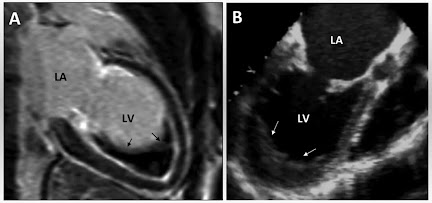
  1. Hypertrophy of cardiac muscles, e.g. systemic hypertension, aortic valve disease (stenosis and insufficiency), mitral insufficiency.
  2. Hypertrophy of smooth muscles, e.g. cardiac achalasia (in oesophagus), pyloric stenosis (in stomach), intestinal strictures, muscular arteries in hypertension.
  3. Hypertrophy of skeletal muscles, e.g. athletes and manual labourers.
  4. Compensatory hypertrophy after removal of contralateral organ, e.g. nephrectomy, adrenal hyperplasia.

Cardiac hypertrophy, adaptation, aasgaduli, pathology
Cardiac Hypertrophy


MORPHOLOGICAL FEATURES – The affected organ is enlarged and heavy. There is enlargement of muscles fibres as well as of nuclei. At ultra-structural level, there is increased synthesis of DNA and RNA, protein and increased number of organelles such as mitochondria, endoplasmic reticulum and myofibrils.


Similar Posts -

Infarction

Infarction

Definition – A necrotic area is called infarct and the process of infarct formation is called infarction.
Causes – Ischaemic necrosis (most common), stagnant hypoxia (venous obstruction), thrombosis, embolism, narrowing of the coronary artery (atherosclerosis) etc.
Types of infarcts –
1. According to colour …
            a. Pale or anemic infarct – It is due to arterial occlusion, commonly seen in compact organ like heart, kidney and spleen.
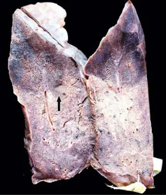
            b. Red or haemorrhagic infarct – It is due to obstruction of the artery, commonly seen in soft loose tissue e.g. lungs and intestine.

Infarct, haemorrhagic infarct, aasgaduli, pathology
Haemorrhagic Infarct of the Lung

2. According to the age … Recent and old.
3. According to the presence or absence of the infection…
            a. Bland – Free from bacterial contamination
            b. Infected – Infected with bacterial contamination.
4. According to the type of action…
            a. Bacteriostatic – It inhibit growth of bacteria e.g. tetracycline, erythromycin, sulfonamide etc.
            b. Bactericidal – It kills bacteria e.g. refampicin, penicillin etc.
5. According to the spectrum of activity… narrow and broad spectrum.
PATHOGENESIS –
  • Localized hyperaemia or congestion may occur due to obstruction of blood supply.
  • Within few hours edema and haemorrhage may occur.
  • Cellular change such as cloudy swelling appear early, necrosis occur within 12-15 hours.
  • Progressive proteolysis of necrotic tissue and there is lysis of red cells.
  • Acute inflammatory reactions appear in surrounding tissue.
  • Blood pigments like haematoidin and haemosiderin deposit in the infarct.
  • Following this granulation tissue starts to appears at the margin, finally infarct is replaced by collagen fibers and a scar is formed.

GROSSLY – Infarcts of solid arms are usually wedge shaped with apex at the obstructed artery and base of the surface of the organ. Most infarcts become pale due to breakdown of red cells but pulmonary infarcts never become pale due to extensive blood supply.

            Cerebral infarcts appear with central softening due to liquefaction necrosis (encephalomalacia), recent infarcts are generally slightly elevated over the surface but old infarcts are shrunken and depressed due to scar.

MICROSCOPICALLY – The tissue of affected area shows coagulation necrosis but in cerebral infarcts there is liquefaction necrosis. At the periphery of infarcts inflammatory reaction is noted. Finally the infarct is replaced by scar, but in cerebral infarcts there is gliosis.


Infarct, haemorrhagic infarct, aasgaduli, pathology
Infarcts of most commonly affected organ


Similar Posts -

Haemorrhage

Haemorrhage

Definition – Haemorrhage is the escape of blood from a blood vessel.
HAEMATOMA – Extravasation of blood into the tissue with resultant swelling is known as haematoma.
ECCHYMOSIS – Large extravasations of blood into the skin and mucous membranes are called ecchymosis.
PURPURA – Small areas of haemorrhage (up to 1 cm) into the skin and mucous membrane called purpura.
PETECHIAE – Minute pinhead size haemorrhages on the skin are called petechiae.
FORMS –
  1. Epistaxis  (nosebleed)
  2. Haemoptysis (coughing up blood)
  3. Haematemesis (vomiting blood)
  4. Haematuria (blood in urine)
  5. Melena (dark stool)
  6. Polymenorrhea (short menstrual cycle <21 days)
  7. Menorrhagia (heavy periods)
  8. Postpartum haemorrhage (excessive bleeding after childbirth)
  9. Antepartum haemorrhage (genital bleeding during pregnancy, third trimester to till birth or delivery)

Classification –
1. According to the nature or type … (a) External or visible haemorrhage (b) Internal or concealed haemorrhage.

2. According to the timing …
            a. Primary haemorrhage – When haemorrhage occurs just after injury of vessels.
            b. Reactionary haemorrhage – When bleeding within 24 hours (usually 4-6 hours) causing slipping of ligature, dislodgement of clot.
            c. Secondary haemorrhage – When haemorrhage occurs after 7-14 days causing surgery, sloughing of vessels due to infection.

3. According to the source…
            a. Arterial haemorrhage – When bleeding occurs due to rupture of artery. Blood is bright red in colour and emitted as spurting jet.
            b. Venous haemorrhage – When bleeding occurs due to rupture of veins. Blood is dark red in colour and emitted steady and copious flow.
            c. Capillary haemorrhage – When bleeding occurs due to rupture of capillaries. Blood is bright red in colour and emitted rapid oozing.

4. According to the duration … (a) Acute haemorrhage (b) Chronic haemorrhage

5. According to the type of intervention… (a) Surgical (b) Non-surgical haemorrhage

 Effects of Haemorrhage –
  • 33% of total blood, sudden loss may cause death.
  • 50% of total blood, loss over the period of 24 hours may not necessary fatal.
  • Chronic haemorrhage generally produce iron deficiency anaemia while occur acute haemorrhage may cause hypovolaemic shock.

Etiology –
  1. Trauma to the vessel wall, e.g. penetrating wound in the heart or great vessels.
  2. Spontaneous haemorrhage, e.g. rupture of an aneurism, septicaemia, bleeding diathesis, acute leukaemias, pernicious anaemia, scurvy.
  3. Inflammatory lesions of the vessel wall, e.g. bleeding from chronic peptic ulcer, typhoid ulcers, traversing a tuberculous cavity in the lung, syphilitic involvement of the aorta, polyarteritis nodosa.
  4. Neoplastic invasion, e.g. vascular invasion in carcinoma of the tongue.
  5. Vascular disease, e.g. atherosclerosis.
  6. Elevated pressure within the vessels, e.g. cerebral and retinal haemorrhage in systemic hypertension, severe haemorrhage from varicose veins due to high blood pressure in the veins of legs or oesophagus.

Necrosis

Necrosis

NECROSIS – Pathological cell or cells death in living body due to injury.
NECROBIOSIS – It is a physiological cell death e.g. desquamation of surface epithelium.
APOPTOSIS – It is a programmed cell death, it may be pathological or physiological. In this process the nucleus is condensed the cell is divided into small membrane bounded bodies called apoptotic bodies. Eventually the apoptotic bodies are engulfed by phagocytes.
Causes –
1. Hypoxia (lack of O2 or ischaemia) – disturbed blood supply.
2. Physical agents – Excessive heat and cold etc.
3. Chemical agents – Strong acids and alkaloids etc.
4. Immunological reactions
General features –
1. The cytoplasm is homogeneous or granular and eosinophilic.
2. The cell cytoplasm may show vacuolation.
3. The nucleus shows three types of changes …
            a. Pyknosis – It is a initial change, in which nuclear material is condensed.
            b. Karyorrhexis – The nucleus is divided into small fragments.
            c. Karyolysis – Finally the nucleus is dissolved out and disappeared.
Types of necrosis –
1. Coagulation
2. Liquefaction or colliquative necrosis
3. Caseous necrosis
1. Coagulation necrosis – It is the most common type of necrosis. It is more often caused by ischaemia and less often caused by bacterial and chemical agents. It is commonly seen in heart, kidney and spleen.
GROSSLY – The affected part is pale, firm, swollen then become yellowish, softer and shrunken.
MICROSCOPICALLY – The necrosed cells are swollen and cytoplasm is eosinophilic with nuclear changes (Pyknosis, Karyorrhexis and Karyolysis) but detail is lost (due to proteolytic enzymes which release from lysosomes) but cell membrane remain intact for days or weeks giving “tomb stone” appearance, finally dead cells are phagocytose remain cell debris behind.

Necrosis, Apoptosis, aasgaduli, Pathology
Contrasting features of necrosis and apoptosis

2. Liquefaction or colliquative necrosis – It is caused by ischaemia, bacterial and fungal infection. The affected part is soft then liquefied later a cyst is form containing necrotic cell debris and macrophages. This type of necrosis is commonly seen in brain. Abscess is also an example of liquefaction necrosis.
Apoptosis, Necrosis, aasgaduli, pathology

3. Caseous necrosis – In this type of necrosis the affected area is converted into yellowish and granular cheese like material. It is commonly seen in the central part of tubercle in tuberculosis.
            Caseous necrosis seen centrally which is surrounded by epithelial cells which are covered by giant cells and loose connective tissue and these all material are covered by connective tissue.


Similar Posts -

Gangrene

Gangrene

Massive cell death of a part of living tissue superadded putrefaction due to saprophytic bacteria.


Classification 
1. Dry gangrene
2. Wet or moist gangrene
3. Gas gangrene

1. Dry Gangrene : This type of gangrene is seen in that part of the body there is lack of water quantity like - hands and feet.

Causes : 
  1. Gradual vascular obstruction as seen in senile gangrene, diabetic gangrene.
  2. Sudden vascular obstruction as seen in thrombosis, embolism and ligation.
  3. Extreme cold (frost bite).
  4. Strong acid, coagulate the fluid of the tissue so produce dry gangrene while strong alkalies liquefies the tissue so produce the moist gangrene.
Features of dry gangrene : 
  1. This type of gangrene is seen in the body there is lack of water like hands and feet.
  2. This type of gangrene generally starts in distal part (great toe) and spread toward the heart.
  3. Growth rate of this type gangrene is slow.
  4. The affected part is initially waxy then becomes dry and shrunken then it become yellowish blue then finally dark blackish due to pigmentary changes ( The stagnant blood RBCs are starts to breakdown and Hb release their iron which combines with hydrogen sulfide that produced by the bacteria and form iron sulfide, which stains the tissue black).
  5. There is found a red line between healthy and dead tissues which is called 'line of demarcation' due to inflammatory reactions, noxious substances produce by necrotic cells.
  6. There are necrotic features are also present.
  • Senile Gangrene : This type of gangrene is seen in old age people due to arteriosclerosis but finally thrombosis is responsible for the occlusion of artery.
  • Diabetic Gangrene : This type of gangrene is due to atheromatous plague in the artery. bacterial growth is favored due to high percent of sugar in red tissue.

Dry gangrene, gangrene, aasgaduli, pathology
Dry gangrene

2. Moist Gangrene : This type of gangrene is seen in the body where, there is present abundant quantity of water like oral cavity, intestine, vulva etc.

Causes : This type of gangrene is heavy develops due to -
  1. Blockage of venous and arterial flow as seen in strangulated hernia, intussusception, volvulus etc.
  2. Burn also produce moist gangrene.
  3. Bed-sore is an example of pressure gangrene may cause occlusion of vessels followed by necrosis and gangrene. It is seen in old age, chronic illness and injury of spinal cord.
Features of moist gangrene : 
  1. This type of gangrene is seen at the area of abundant water.
  2. The affected part is cold and pulseless.
  3. The color of affected part is bluish to green and black due iron sulfide.
  4. There is absent line of demarcation.
  5. The rate of spread is rapid.
  6. Growth of bacteria also rapid due to excessive fluid concentration. Toxins of bacteria are absorbed and produce severe toxaemia and even death.

Moist gangrene, wet gangrene, gangrene, aasgaduli, pathology
Moist or wet gangrene

3. Gas or Infective Gangrene : This type of gangrene is caused by the infection of anaerobic bacteria it is seen in wounds.

Causes : This type of gangrene is produced by following clostridia species -
A). Saccharolytics
  • Clostridium welchii (80%)
  • Clostridium septicum
B). Proteolytics
  • Clostridium histolyticum
  • Clostridium sporogenes
Dry gangrene, wet gangrene, gangrene, aasgaduli, pathology
Contrasting features


Pathogenesis
  1. The infection of anaerobic bacteria, laceration and crush injury is essential. The organism don't grow with healthy tissue.
  2. Haemorrhage and blood clot help the infection specially by calcium supply.
  3. Contaminated soil, supplies silica and calcium to the wounds is also help in infection.
  4. Dirty splits and fragments causes laceration and contamination of wounds.
  5. Circulatory obstruction caused by the occlusion or damage of the artery. pressure on vessels by tourniquet or tight bandage are contributory factors.
  6. Inadequate drainage and exudate and favours the spread of infection.
Infection of damage tissue by Streptococci, E. Coli, Staphylococci etc. produce anaerobic atmosphere by loosing oxygen of the tissue that's by giving the chance when anaerobic bacteria to grow. 
        The saccharolytic group of organism breaks the glycogen into carbon dioxide, hydrogen and lactic acid. Excessive production of lactic acid  stop the growth of saccharolytic organism at the same time proteolytic group of organism is starts to growth and liberate proteinase and forms the amino acid in the tissue. This amino acid breakdown into ammonia which neutralized lactic acid. At the same way the cycle repeat again and again.
        The liberation of toxins in blood causing increase blood pressure. This toxins also damage the endocardium causing sudden heart failure. The gas gangrene producing bacteria produce enzymes hyaluronidase, proteinase and collagenase which helps in spread of lesion.

Morphological features - The affected part is swollen and crepitant on palpation due to accumulation of gas in the tissue. The color of skin is affected part is yellowish green or black. On microscopic examination  the causative organism are seen, the muscle fibers lose striation hyalinized and disintegrated.

Atrophy

ATROPHY
Definition – Reduction of the number and size of parenchymal cells of a normal organ or its parts called atrophy.
HYPOPLASIA – Incomplete developments of a tissue or organ.
APLASIA – Defective development or congenital absence of a tissue or organ.
Causes – It may be physiological or pathological…
PHYSIOLOGICAL ATROPHY –
  • Atrophy of lymphoid tissue with age.
  • Atrophy of thymus in adult life.
  • Atrophy of gonads after menopause.
  • Atrophy of brain with ageing.
  • Osteoporosis with reduction in size of bony trabeculae due to ageing.

PATHOLOGICAL ATROPHY –
  1. Starvation atrophy – This type of atrophy is due to lack of food and using stored food. It is seen in cancer and severely ill patients.
  2. Ischaemic atrophy – Gradual diminution of blood supply due to atherosclerosis may result in shrinkage of the affected organ e.g. small atrophic kidney and atrophy of the brain due to atherosclerosis.
  3. Disuse atrophy – Prolonged diminished functional activity may cause atrophy e.g. wasting of muscles of limb immobilized in cast and atrophy of the pancreas in obstruction of pancreatic duct.
  4. Neuropathic atrophy – Interruption in nerve supply leads to wasting of muscles e.g. poliomyelitis, motor neuron disease, nerve section.
  5. Endocrine atrophy – Loss of endocrine regulatory mechanism results in related tissue atrophy e.g. hypopituitarism may lead to atrophy of thyroid, adrenal and gonads. Hypothyroidism may cause atrophy of skin and its adnexal structures.
  6. Pressure atrophy – Prolonged pressure from benign tumours or cyst or aneurysm may cause compression and atrophy of the tissue e.g. erosion of the spine by tumour in nerve root, erosion of the skull by meningioma, erosion of the sternum by aneurysm of arch of aorta.
  7. Idiopathic atrophy – Atrophy without known cause e.g. myopathies, testicular atrophy.
Atrophy, adaptive disorders, aasgaduli, pathology
Adaptive disorders of growth


MORPHOLOGICAL FEATURES – Affected organ is small due cell size decrease, often shrunken due to reduction in cell organelles chiefly mitochondria, myofilaments and endoplasmic reticulum. There is increase number of autophagic vacuoles containing cell debris, these vacuoles may persists to form ‘residual bodies’ in the cell cytoplasm e.g. lipofuscin pigment granules in brown atrophy.


Endomyocardial Fibrosis