Showing posts with label Physiology. Show all posts

Body fluids

Body Fluids

Body fluids are essential for maintaining physiological balance and enabling various biochemical processes within the body. These fluids exist within cells, surrounding tissues, and in specialized compartments, playing a critical role in transportation, lubrication, temperature regulation, and waste elimination.

Nucleus

Name :- Nucleus (Nucleus was first described by Robert Brown in 1831, and it's material stained by the basic dyes was given the name chromatin by Flemming.)


Properties :

  • It is the largest cellular organelle.
  • Generally located in the center of the cell but it may vary in some cells.
  • It's diameter is about 10µ to 22µ.
  • Mostly it is spherical in shape but it may vary in some cells.
  • Nucleus occupies about 10% of total volume of the cell.
  • It is present in all the cells in the body except RBCs.
  • The cells with nucleus called eukaryotes and without nucleus called prokaryotes.
  • Presence of nucleus is necessary for cell division.
  • Most of the cells have only one nucleus called uninucleated cells.
  • Few types of cells like skeletal muscle have many nuclei called multinucleated cells.
Nucleus


Structure Of Nucleus :
  1. Nuclear membrane
  2. Major components like nucleoplasm, chromatin, chromosomes and nucleolus.
 1. Nuclear Membrane (NM) :

  • NM is double layered membrane called nuclear envelope, separates the nucleus from the cytoplasm.
  • NM is porous in nature, which allows the nucleoplasm to communicate with the cytoplasm.
  • Pores (diameter - 80-100 nm) of the NM are lined by protein molecules (after attachment of protein molecules, pores diameter may decrease 7-9 nm).
  • Small molecules and ions move through the pores by passive diffusion, while large molecules such as RNAs and proteins pass through the selectively active transport.
  • Outer membrane of NM is continue with membrane of endoplasmic reticulum (ER).
  • The space between two layered of NM is continue with the lumen of the ER.
2. Major Components : Such as nucleoplasm, chromatin, chromosomes and nucleolus ....

Nucleoplasm :
  • Nucleoplasm is a highly viscous fluid similar to cytoplasm.
  • It surrounds chromatin and nucleolus.
  • It contains nuclear matrix (dense fibrillar network of protein) and many substances such as nucleotides, enzymes and hyaloplasm (soluble liquid part of nucleoplasm).
  • Nuclear matrix forms the structural framework for organizing chromatin.
Chromatin : 
  • Chromatin is a thread like material referred as DNA-histone complex or complex of DNA, proteins (histones) and some RNA. (A chain of nucleosomes)
  • DNA is a double helix which wraps around the central core of eight histone molecules to form the fundamental packing unit of chromatin called nucleosome. (Eight histones wrapped by double helix DNA)
  • Nucleosomes are packed together tightly with the help of histone molecule to form a chromatin fiber.
  • Just before cell division, the chromatin condenses to form a pair of chromatids that constitutes a chromosome.
  • In the cell that are not dividing, the chromatin appears as a diffuse granular mass.

Nucleus


Chromosomes : 
  • Chromosome is the rod-shaped nuclear structure that carries a complete blueprint of all the hereditary characteristics of that species.
  • Each chromosome is a long molecule of DNA (contains many genes) that is coiled together with several proteins.
  • Normally, the chromosomes are not visible in the nucleus under microscope while it visible (because DNA more tightly packed just before cell division) only during cell division.
  • All the dividing cells of the body except reproductive cells contain 23 pairs of chromosomes.
  • Each pair consists of one chromosome inherited from father and one from mother.
  • The cells with 23 pairs of chromosomes are called diploid cells. The cells with 23 single chromosomes called haploid cells like reproductive cells also called for this gametes or sex cells
Nucleolus (pleural - nucleoli) :
  • Nucleolus is a small, round granular structure of the nucleus.
  • Each nucleus contain one or more nucleoli.
  • Nucleoli are quite prominent in cells that synthesize large amount of protein like muscle and liver cells.
  • Nucleoli disperse and disappear during cell division and reorganize once new cells are formed.
  • The nucleolus contains RNA and some proteins which are similar to those found in ribosomes.
  • The RNA is synthesized by five different pairs of chromosomes and stored in nucleolus. Later, it condensed to form sub-units of ribosomes. All the sub-units are transported from nucleolus to cytoplasm through pores of the nuclear membrane.
Functions Of Nucleus :
  1. Control of the all activities of the cell such as metabolism, protein synthesis, growth and reproduction (cell division).
  2. Synthesis of RNA.
  3. Formation of sub-units of ribosomes.
  4. Sending genetic instruction to the cytoplasm for protein synthesis through messenger RNA (mRNA).
  5. Control of the cell division through genes.
  6. Storage of hereditary information (in genes) and transformation of this information from one generation of the species to the next.

Similar Posts :

Golgi apparatus

Name :- Golgi apparatus, Golgi body, Golgi complex, Post office of the cell, Shipping department of the cell (Camillo Golgi (1898) first discovered, hence the name is Golgi).

Properties :
  • It is an membrane bound organelle situated near the nucleus.
  • It is present in all cells except RBCs.
  • Usually, each cell has one Golgi apparatus.
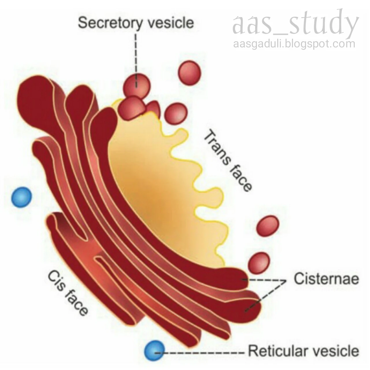
  • Each Golgi apparatus consists of 5-8 flattened membranous sacs called cisternae (0.5-1.0 micrometer).
  • Golgi apparatus has two ends or faces, namely cis face (situated near the endoplasmic reticulum) and trans face (situated near the cell membrane).
Functions :
Major functions of Golgi apparatus are processing, packing, labeling and delivery of proteins and lipids to different part of the cell or outside of the cell.
  1. Processing :- glycoproteins and lipids are modified here.
  2. Packaging :- modified materials are packed in the form of secretory granules, secretory vesicles and lysosomes.
  3. Labeling and delivery :- Golgi apparatus sort out the processed materials and labels them (such as phosphate group) then distribute in or out side of the cell according to demand.

Golgi apparatus


Similar Posts :-

Functions of Lipid Layer in cell membrane

Functions of Lipid layer in cell membrane

  1. Semipermeability : Lipid layer act as a semipermeable membrane which allow only fat-soluble substances through it.
  2. Permeability : Lipid  layer act as a permeable membrane which allow Oxygen, Carbon dioxide and alcohol.
  3. Prevention : The water soluble substances like Glucose, Urea and Electrolytes cannot through this layer. 


Source : Essential MEDICAL PHYSIOLOGY

Functions of Carbohydrate in cell membrane

Functions of Carbohydrate in cell membrane

  1. Carbohydrate molecules are negatively charged and do not permit the negatively charged substances to move in and out of the cell.
  2. Glycocalyx from the neighboring cells helps in the tight fixation of cell with one another.
  3. Some carbohydrate molecules function as the receptors for some hormones.


Source : Essential MEDICAL PHYSIOLOGY

Functions of protein in cell mambrane

Functions of Protein in cell membrane


  1. Integral proteins provide the structural integrity of the cell membrane.
  2. Channel proteins help in the diffusion of water soluble substances like glucose and electrolytes.
  3. Carrier or  transport proteins help in the transport of substances across the cell mambrane with the help of active and passive transport.
  4. Some carrier proteins act as a pump, by which ions are transported actively across the cell membrane.
  5. Receptor proteins serve  as the receptor sites for hormones and neurotransmitters.
  6. Some protein molecules forms the enzymes and control chemical (metabolic) reactions within the cell membrane.
  7. Some proteins act as a antigens and induce the the process of antibody formation.
  8. Cell adhesion molecules or the integral proteins are responsible for the attachment of cells to their neighbors or to basal lamina.


Colour blindness

Colour blindness

It is a condition in which a person is unable to identify one or more colours.

Incidence

Male : it present about in 8% male.
Female : it present about 0.4% female.

Causes

  • Injury to retina.
  • Disease of retina.
  • Inherited disease

Types

  1. Monochromatism
  2. Dichromatism
  3. Trichromatism

Monochromatism

It is a rare condition in which a person is unable to identity any colour vision is similar to black and white photograph and everything looks grey.

Types


  1. Rod monochromatism
  2. Cone menochromatism

Dichromatism


In this condition person can identify only two basic colours.

Types

  1. Protanopia / Primary dichromatism - Unable to identify red colour.
  2. Deuteranopia / Secondary dichromatism - Unable to identify green colour.
  3. Tritanopia / Tertiary diachromatism - Unable to identify blue colour.

Trichromatism

In this condition person can identify all three in colours but the intensity of one of primary colour can not identify properly.

Types

  1. Protanomaly / Primary trichromatism - Intensity for red colour is less
  2. Deuteranomaly / Secondary trichromatism - Intensity for green colour is less.
  3. Tritalomaly / Tertiary trichromatism - Intensity for blue colour is less.

Regulation of blood sugar level

Regulation of blood sugar level

Glucose is the ultimate source of energy to our body so amount of sugar in blood should be maintained in normal range.
Any condition when increase blood sugar level is known as hyperglycemia ( above 140 mg/dl in fasting condition ), which may result from disease like diabetes mellitus.
Any condition when blood sugar level decrease  called hypoglycemia ( below 55 mg/dl in fasting condition ), which may cause multiple symptoms and even may cause death.

Control blood sugar level through hormones

1. Insulin

It secrets from beta cells of islets of pancreas.
It decrease blood sugar level by...

  • Gluconeogenesis : it decrease gluconeogenesis.
  • Glycogenesis : it increase the glycogenesis.


2. Adrenaline

It secret from adrenal glands.
It increases the blood sugar level by...

  • Gluconeogenesis : it increase gluconeogenesis.
  • Glycogenesis : it decrease glycogenesis.


3. Nor adrenaline

It secret from adrenal gland.
It increase blood sugar level by...

  • Gluconeogenesis : it increase gluconeogenesis.
  • Glycogenesis : it decrease glycogenesis.


 4. Glucagon

It secret from alpha cells of islets if pancreas.
It increase blood sugar level by...

  • Gluconeogenesis : it increase gluconeogenesis.
  • Glycogenesis : it decrease glycogenesis.


5. Cortisol

It release from adrenal glands.
It increases the blood sugar level by...

  • Gluconeogenesis : it increase gluconeogenesis.
  • Glycogenesis : it decrease glycogenesis.


6. Thyroxin

It secret from thyroid gland.
It increases the blood sugar level by...

  • Gluconeogenesis : it increase gluconeogenesis.
  • Glycogenesis : it decrease glycogenesis.


7. Growth hormone

It secret from anterior pituitary gland.
It increases the blood sugar level by...

  • Gluconeogenesis : it increase gluconeogenesis.
  • Glycogenesis : it decrease glycogenesis.


Hyperglycemia ( Diabetes Mellitus )

Hyperglycemia ( Diabetes Mellitus )

It is the condition which is characterized by increase level of sugar in blood that is...
1. Fasting level is more then 140 mg /dl
2. After meal the blood sugar level is more than 200 mg /dl

While normal blood sugar level is
In fasting condition : 70 - 110 mg/dl
After meal : 140 mg/dl

Symptoms

  • Hyperglycemia ( increase blood sugar level )
  • Glucosuria ( presence of sugar in urine )
  • Polyuria ( frequent urination )
  • Polydipsia ( increase thirst )
  • Muscles wasting ( muscle dysfunction )

 Complications

  • Development of atheroma ( degeneration of wall of the arteries due to fatty deposit and scar tissue )
  • Nephropathy ( kidney disease or damage )
  • Retinopathy ( any damage of retina of the eye )

Types of diabetes mellitus

1. IDDM ( Insulin dependent diabetes mellitus )

It occurs due to the absence of insulin or non functioning of beta cells.

2. Non IDDM ( Non insulin dependent diabetes mellitus )

In this disease insulin are present normally but there is defect between receptor and target cells.

Hypoglycemia

Hypoglycemia

It is the condition which is characterized by decrease amount of sugar in blood that is...
less than 55 mg/dl

While normally blood sugar level is...
In fasting condition : 70 - 110 mg/dl
After meal : 140 mg/dl

Symptoms

  • Sweating
  • Palpitation
  • Giddiness ( a sensation of whirling and tendency to fall )
  • Confusion
  • Convulsion ( a sudden violent movement of the body that you can not control )
  • Comma ( death of brain )

Cerebro spinal fluid (CSF)

Cerebrospinal fluid or CSF

CSF is the modified colour less tissue fluid. CSF present in ventricular system of brain and sub arachnoid space around spinal cord and brain.

Formation

Maximum part of CSF is formed by choroid plexus of lateral ventricle, and lesser part of CSF  is formed by third and fourth ventricles. Some part of CSF  is also formed by capillaries present surface of spinal cord.

Quantity                 : 150 ml
Rate of formation : 0.3ml / min
Reaction                 : Alkaline
Specific gravity     : 1.005
Coagulation           : It does not coagulate in standing

Pressure exerted  in man by CSF, varies in different position...
  • Lateral recumbent position : 10 - 18 cm of water
  • Lying position                         : 13 cm of water
  • Sitting position                        : 30 cm of water

Composition of CSF

1. Water - 99.13 %

2. Solids - 0.87%

A. Organic substances

  1. Proteins          : 20-30 mg / dl
  2. Amino acids   : 10 -30 mg / dl
  3. Sugar              : 50 - 80 mg / dl
  4. Cholesterol     : 0.06 - 0.22 mg /dl
  5. Urea                : 10 - 30 mg / dl
  6. Uric acid         : 0.5 - 2.2 mg / dl
  7. Creatinine       : 0.5 - 2.2 mg / dl
  8. Lactic acid      : 8.27 mg / dl

 B. Inorganic substances

  1. Sodium         : 350 mg / dl
  2. Calcium
  3. Potassium     : 12 mg / dl
  4. Magnesium   : 3 - 3.6 mg / dl
  5. Chlorides      : 700 - 750 mg / dl
  6. Phosphate     : 1.8 mg / dl
  7. Bicarbonates : 40 - 60 mg / dl
  8. Sulfates         : 0.6 mg / dl
CSF also contain some lymphocytes ( 6 / cu mm ). These lymphocytes are not present when it secrets from ventricles. Lymphocytes are added when CSF flows in spinal cord.

Cerebrospinal fluid, CSF, Aas, Alfaaz-e-aas
Composition of CSF


Absorption of CSF

  1. It absorbed mainly through arachnoid villi and it drain into cranial venous sinuses.
  2. It also absorbed by lymphatic present around 2nd, 7th and 8th cranial nerves.
  3. It also absorbed by veins around spinal nerves. 

Circulation

  • CSF passes from lateral ventricles to third ventricle by the inter ventricular foramen or foramen of monro.
  • It passes from third to fourth ventricle by the Aqueductus Sylvius.
  • It  enters from fourth ventricle to cisterna magna and cisterna lateralis through foramen of magendie ( central opening ) and foramen of luschka ( lateral opening ).
  • From cisterna magna and cisterna lateralis, CSF  circulates through subarachnoid space and cerebral hemispheres. It  flows also into the central canal of spinal cord.
CSF, Circulation of Cerebrospinal fluid, Aas, Alfaaz-e-aas
Circulation of CSF

Functions

  1. Protection
  2. Nutrition
  3. Excretion

Applied physiology

1. For diagnosis of disease, bio-chemical examination of CSF.
2. Any obstruction to flow of CSF in ventricle it causes...
  • Increase intra cranial pressure in adults.
  • Hydrocephalus in children's.
3. CSF is obtained by...
  • Lumbar puncture - between 3rd and 4th lumbar spine.
  • Cisternal puncture.
  • Ventricular puncture.


Images copied from Physiology Sembulingum book

Parkinson's disease or Parkinsonism

Parkinson's disease or Parkinsonism

Parkinson's disease was discovered by James Parkinson. Name of this disease are given on the name of it's discoverer. This disease are also called Paralysis Agitates.
Parkinsons disease occurs due to damage of basal ganglia. It is mostly due to the destruction of substantial nigra and nigro-strial pathway, which has dopaminergic fibers.

Causes

  1. Injury to basal ganglia
  2. Cerebral arteriosclerosis
  3. Viral infection of brain like encephalitis

Symptoms

  1. Rigidity of muscles
  2. Tremors
  3. Akinesia or hyperkinesia
  4. Abnormal gait
  5. Emotional changes

Treatment

Parkonism disease is due to the damage of dopaminergic fibers, so it's treated by dopamine.

Organ of corti

Organ of CORTI

Organ of corti is the organ receptor for hearing. It is the neuro epithelial structure in cochlea. It rest's upon the  lip of osseous spiral lamina and the basilar membrane. it extent's through the cochlear duct, except for a short distance on either end.

The organ of corti is made up of sensory elements called the hear cells. All the cells of organ of corti are arranged in order from center to towards the periphery of cochlea.

  1. Border cells
  2. Inner hair cells
  3. Inner hair phalange cells
  4. Inner pillar cells
  5. Outer pillar cells
  6. Outer phalange cells
  7. Outer hair cells
  8. Cells of Hansen
  9. Cells of Dandies
  10. Tectorial membrane and lamina reticularis.

Loss of speech or Aphasia

Loss of speech or Aphasia

Aphasia is defined as the loss or impairment of speech due to the brain damage. It is an acquired disorder and it is distinct from developmental disorder of speech.
Aphasia is not due to the paralysis of muscles of articulation.

Cause

Damage to the speech centers, which are situated in cerebral cortex.

Reasons of the damage of speech center

  • Stroke
  • Head injury
  • Cerebral tumors
  • Brain infections

Types of Aphasia

  1. Broca's aphasia
  2. Wernicke's aphasia
  3. Glossal aphasia
  4. Anomic aphasia

Digestion of lipid

Digestion of lipid

Definition of digestion

Brocken down of big food particles into small food particles, which can be absorbed and use as a nutrients for the body.

Classification of lipid

1. Simp;e fat -

a. Triglyceride / Neutral fat 
b.  cholesterol .

2. Compound fat -

a. Phospholipid
b. Glycolipid
c. Lipoprotein

3. Derived fat -

a. Prostaglandin
b. Leuketrin
  • We are lipid intake with food in the form of triglyceride mostly while some parts of cholesterol and cholesterol ester.
  • The enzyme which help in digestion of lipid called lipolytic  enzyme.  


Lipid, Digestion of lipid, Aas, Meranazarya, Physiology

Digestion of lipid in mouth

Saliva contain a lipolytic enzyme called lingual lipase, but this enzyme not act on food in mouth and engulfed with food.

Digestion of lipid in stomach

Gastric juice contain a lipolytic enzyme called gastric lipase, which act on triglyceride and converts into fatty acids and glycerol.

Digestion of lipid in small intestine

Intestine have two juices...
1. Pancreatic juice
2. Intestinal juice / succus entericus

Pancreatic juice

It contain enzymes...
1. Pancreatic lipase - it act on triglyceride and convert into fatty acids.

2. Cholesterol ester hydrolase - it act on cholesterol ester  and convert  into free cholesterol.

3. Phospholipase - it act on phospholipid and converts into free phospholipid and fatty acid.

4. Bile salt activated lipase - it activate bile for emulsification of fat. This enzyme act firstly and activated bile for emulsification of fat like fatty acid and cholesterol  because these are insoluble in water where as phospholipid are soluble in water.    

Intestinal juice / succus entericus

It contain only a lipolytic enzyme called intestinal lipase (weak enzyme) , which act on triglyceride and convert into fatty acid and glycerol ( it's function is negligible ).

Absorption

Lipid absorbed from small intestine.

  • Less then 12 carbon fatty acid absorb in the form of micelles.
  • More then 12 carbon fatty acids absorb in the form of chylomicrons.

Digestion of protein

Digestion of protein


Definition

Broken down of big food particles into small food particles by the chemical process, which can be absorbed and use as a nutrients.
  • The enzyme which help in protein digestion called proteolytic enzyme.

Protein, Aas, Digestion of protein
Enzymes of protein digestion


Digestion of protein in mouth

Saliva secrets in the mouth which don't have any proteolytic enzymes, so protein digestion not occur in the mouth.

Protein digestion in stomach

Gastric juice contain a proteolytic enzyme called pepsin. Pepsin act on protein and converts into protease, peptone and polypeptide. pepsin act in highly acidic medium.

Digestion of protein in small intestine

Small intestine contain two juices...
1. Pancreatic juice
2. Succus entericus

Pancreatic juice

Pancreatic juice have carboxy peptidase and endo-peptidase ( trypsin and chymotrypsin ) enzymes, these enzyme are proteolytic enzyme.
  • Trypsin act on protein and convert into di-peptide.
  • Chymotrypsin act on peptone and convert into tri-peptides and poly-peptide.
  • Carboxy-peptidase act on dipeptide, tri-peptide and poly-peptide and converts into amino-acids.

Succus entericus

It has many enzymes...
  • Mono-peptidase - it act on mono-peptide and convert into amino-acids.
  • Di-peptidase - it act on dipeptide and convert into amino-acids.
  • Tri-peptidase - it act on tri-peptide and convert into amino-acids.
  • Poly-peptidase - it act on poly-peptide and convert into amino-acids.
  • Protein absorbed with sodium in the form of amino-acids from small intestine.
  • Protein fastly absorbed and duodenum and jejunum and slowly in the ilium.


Digestion of carbohydrate

Digestion of carbohydrate

Definition

Brockendown of  big food particles into small food particles by the chemical process, which can be absorb and use as a nutrients.

The enzyme which help in digestion of carbohydrate called amylolytic enzyme.

Classification of carbohydrates


1. Monosaccharide - Hexoxes ( glucose ) , Pantoses.
2. Disaccharide - Sucrose ( Glucose + Fructose ), Lactose ( Glucose + Galactose )
3. Polysaccharide - Starch, Glycogen, Amylose.


Digestion of carbohydrates, Digestion
Enzymes of digestion of carbohydrate

Digestion in mouth

Mouth secrets saliva which contain salivary amylase/ ptylin /ptyalin. This enzyme act as amylolytic enzyme. This enzyme acts on starch and converts into dextrose and maltose.
Ptyalin inactive in highly acidic medium, so when food reach in stomach then ptyalin is inactive due to increase acidity.

Digestion in stomach  

Gastric juice contain a enzyme called gastric amylase, which is a very weak enzyme so it's function is negligible in stomach.

Digestion in intestine

Intestine have two juices...
1. Pancreatic juice
2. Succus entericus / Intestinal juice

Pancreatic juice

It contain pancreatic amylase which act on starch and converts into maltose and dextrose.

Intestinal juice

This juice contain many enzymes...
1. Sucrase - it act on sucrose and convert into glucose
2. Maltase - it act on maltose and convert into glucose
3. Lactase - it act on lactose and convert into glucose and galactose
4. Dextrinase - it act on dextrose and convert into glucose

  • Carbohydrate absorb from small intestine in the form of glucose (80%), fructose and galactose (20%). 

Cardiac cycle

Cardiac cycle

Definition

The change occur in heart during one heart beat, this process continue repeated in sequence manor called cardiac cycle.

Duration of cycle


  • Heart rate - 70 - 80 / min.
  • Average - 75 /  min.
  • One heart beat - 60/75 = 0.8 sec.


Each cardiac cycle is starts from spontaneous generation of impulses by SA node, this impulse travel from SA node to AV node through intermodal fibers than bundle of his than right and left bundle branch than purkinge fibers than it distributes all myocardium.

Heart, cardiac cycle, events
Events of cardiac cycle


Events of cardiac cycle

Atrial events ( 0.8 sec )

1. Atrial systole  ( 0.1 sec ).
2. Atrial diastole ( 0.7 sec ).

Ventricular events ( 0.8 sec )

1. Ventricular systole ( 0.3 sec ).
2. Ventricular diastole ( 0.5 sec ).

Ventricular systole ( 0.3 sec )

1. Isovolumetric phase ( 0.05 sec ).
2. Rapid ejection phase ( 0.1 sec ).
3. Reduce ejection phase ( 0.15 sec ).

1. Iso-volumetric phase ( 0.05 sec )


  • Valve - close all valves.
  • Heart sound - 1st heart sound produce.
  • Ejection - not start.
  • Contraction - begins.
  • Volume - same ( not change ).

2. Rapid ejection phase ( 0.1 sec )


  • Valve - semilunar valve are open and atrio-ventricular (AV) valve are remain closed.
  • Heart sound - no.
  • Ejection - rapidly.
  • Contraction - continue.
  • Volume - reduced fastly.

 3. Reduced ejection phase ( 0.15 sec )


  • Valve - semilunar valve are open and AV valve remain closed.
  • Heart sound - no.
  • Ejection - slowly-slowly.
  • Contraction - continue.
  • Volume - reduced.

Ventricular diastole ( 0.5 sec )

1. Proto-diastolic phase ( 0.04 sec ).
2. Iso-volumetric relaxation ( 0.06 sec ).
3. Rapid filling phase ( 0.1 sec ).
4. Slow filling phase ( 0.2 sec ).
5. Last rapid filling phase ( 0.1 sec ).

1. Proto-diastolic phase ( 0.04 sec )


  • Valve - closer of semilunar valve and AV valve are already closed.
  • Heart sound - 2nd heart sound produce.
  • Filling - not starts.
  • Relaxation - ready to beginning.
  • Volume - remain end diastolic volume ( 60ml ).

2. Iso-volumetric relaxation ( 0.06 sec )


  • Valve - all valves are closed.
  • Heart sound - no.
  • Filling - not start.
  • Relaxation - ventricle relaxed.
  • volume - no change.

3. Rapid filling phase ( 0.1 sec )


  • Valve - open AV valves.
  • Heart sound - 3rd heart sound produce.
  • Filling - rapidly
  • Relaxation - already relaxed.
  • Volume - increase ( 70% filling ).

4. Slow filling phase ( 0.2 sec )


  • Valve - AV valve remain open and semilunar valve are closed.
  • Heart sound - no.
  • Filling - slowly-slowly.
  • Relaxation - already relaxed.
  • Volume - ( 20% filling only in this phase ).

5. Last rapid filling phase ( 0.1 sec )


  • Valve - AV already open and semilunar remain closed.
  • Heart sound - 4th heart sound produce.
  • Filling - rapidly.
  • Relaxation - already relaxed .
  • Volume - both chamber complete filled ( 10% filling only in this phase ).

Principles of Anatomy and Physiology by Geraro J. Tortora and Bryan Derrickson


Principles of Anatomy and Physiology by Geraro J. Tortora and Bryan Derrickson


Anatomy and physiology Tartora book pdf
Tartora book pdf


Download book pdf.

About file:-

File type - pdf
File size - 105.01 MB
Pages - 1281
Language - English 

Text book of Medical Physiology by Gyton and Hall


Text book of Medical Physiology by Gyton and Hall


Medical Physiology Gyton book pdf
Gyton book pdf


Download book pdf

About file:-

File type - pdf
File size - 46.58 MB
Pages - 1046
Language - English

Endomyocardial Fibrosis