We offer topic-wise short notes and medical books to support medical students in their studies.
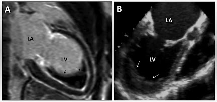
Endomyocardial Fibrosis
Endocardial Fibroelastosis
Endocardial Fibroelastosis
 |
| Image source Google |
Cardiac Amyloidosis
Cardiac Amyloidosis: An In-Depth Analysis
Cardiac amyloidosis is a progressive and life-threatening disorder characterised by the deposition of misfolded amyloid fibrils within the myocardium. This pathological protein accumulation results in increased myocardial stiffness, diastolic dysfunction, and, ultimately, heart failure. Due to its nonspecific clinical manifestations, cardiac amyloidosis often remains undiagnosed until advanced stages, highlighting the necessity for heightened clinical awareness and improved diagnostic methodologies.
 |
| Image source Google |
Dilated Cardiomyopathy
Idiopathic Dilated or Congestive Cardiomyopathy: A Comprehensive Guide
Idiopathic dilated or congestive cardiomyopathy is a heart condition that weakens and enlarges the heart muscle, impairing its ability to pump blood effectively. In simple terms, it’s like the heart becoming overstretched and losing its strength, making it harder for the body to receive the oxygen-rich blood it needs. Globally, idiopathic dilated or congestive cardiomyopathy affects millions, with estimates suggesting it accounts for approximately 25% of all cases of heart failure. Its significance lies in its profound impact on life expectancy and quality of life. Patients with idiopathic dilated or congestive cardiomyopathy often face challenges such as fatigue, shortness of breath, and even sudden cardiac arrest if left untreated. Understanding this disease is crucial for early detection and effective management.
 |
| Image source Google |
 |
| Image source Google |
Obstructive Cardiomyopathy
Obstructive Cardiomyopathy: A Comprehensive Overview
Hypertrophic Obstructive Cardiomyopathy (HOCM) is a subtype of hypertrophic cardiomyopathy characterized by abnormal thickening of the myocardium, predominantly affecting the interventricular septum. This hypertrophy can impede blood flow from the left ventricle into the aorta, posing significant hemodynamic challenges. HOCM is a relatively rare disorder, with a prevalence of approximately 1 in 500 individuals, yet its implications for cardiovascular health are profound. Understanding the condition requires a multidisciplinary approach, encompassing insights from genetics, imaging, and interventional cardiology to ensure accurate diagnosis and effective management.
 |
| Image source Google |
Hypertrophy Of The Heart
HYPERTROPHY OF THE HEART
Definition - Hypertrophy
of the heart is defined as an increase in size and weight of the myocardium. In
which involve predominantly the left or the right or both sides of the heart. It
generally results from increase pressure load while increased volume load
results in hypertrophy with dilatation. It is a compensatory mechanism of the
heart.
Common
causes – (usually causes are
unknown)
- Stretching
of myocardial fibrosis.
- Anoxia
(e.g. in coronary atherosclerosis).
- Certain
hormones (e.g. Catecholamines and pituitary growth hormone).
- Left
ventricular hypertrophy.
Causes of left ventricular
hypertrophy –
- Systemic
hypertension.
- Aortic
stenosis and insufficiency / regurgitation.
- Mitral
insufficiency / regurgitation.
- Coarctation
of the aorta.
- Occlusive
coronary artery disease.
- Congenital
anomalies like septal defects and patent ductus arteriosus.
- Conditions
with increased cardiac output e.g. thyrotoxicosis, anaemia, arteriovenous
fistula.
Causes of right ventricular
hypertrophy –
- Pulmonary
stenosis and regurgitation.
- Tricuspid
regurgitation.
- Mitral
stenosis and regurgitation.
- Chronic
lung diseases (e.g. chronic emphysema, bronchiectasis, pneumoconiosis,
pulmonary vascular disease).
- Hypertrophy
and failure of left ventricle.
Causes of dilatation –
- Valvular
regurgitation.
- Left
to right shunt (e.g. in ventricular septal defect (VSD).
- Conditions
with high cardiac output (e.g. thyrotoxicosis, arteriovenous shunt).
- Myocardial
diseases (e.g. cardiomyopathies, Myocarditis).
- Systemic
hypertension.
Morphological features - Hypertrophy
of myocardium without dilatation called concentric and with dilatation called
eccentric hypertrophy. The weight of the heart is increased above normal
(500 gm). Excessive pericardial fat not indicate true hypertrophy. Thickness of
the left ventricular wall (above 15 mm) is indicates hypertrophy.

Schematic diagram showing transverse section through the ventricles with left ventricular hypertrophy (concentric and eccentric)
GROSSLY -
- In
concentric hypertrophy, the lumen of the chamber is smaller than normal, while
in eccentric hypertrophy the lumen is dilated.
- Papillary
muscles and trabeculae carneae are rounded and enlarged in concentric
hypertrophy, while flattened in eccentric hypertrophy.
MICROSCOPICALLY - There
is increase in size of myocardium. There may be multiple minute foci of
degenerative changes and necrosis in affected myocardium resulting in develop
hypoxia due to inadequate blood supply to increased muscle fibers, while
ventricular hypertrophy is lead to ischaemia.
Similar Posts -
- Systemic hypertension.
- Aortic stenosis and insufficiency / regurgitation.
- Mitral insufficiency / regurgitation.
- Coarctation of the aorta.
- Occlusive coronary artery disease.
- Congenital anomalies like septal defects and patent ductus arteriosus.
- Conditions with increased cardiac output e.g. thyrotoxicosis, anaemia, arteriovenous fistula.
- Pulmonary stenosis and regurgitation.
- Tricuspid regurgitation.
- Mitral stenosis and regurgitation.
- Chronic lung diseases (e.g. chronic emphysema, bronchiectasis, pneumoconiosis, pulmonary vascular disease).
- Hypertrophy and failure of left ventricle.
- Valvular regurgitation.
- Left to right shunt (e.g. in ventricular septal defect (VSD).
- Conditions with high cardiac output (e.g. thyrotoxicosis, arteriovenous shunt).
- Myocardial diseases (e.g. cardiomyopathies, Myocarditis).
- Systemic hypertension.
 |
| Schematic diagram showing transverse section through the ventricles with left ventricular hypertrophy (concentric and eccentric) |
- In concentric hypertrophy, the lumen of the chamber is smaller than normal, while in eccentric hypertrophy the lumen is dilated.
- Papillary muscles and trabeculae carneae are rounded and enlarged in concentric hypertrophy, while flattened in eccentric hypertrophy.
Similar Posts -
Endocarditis
Endocarditis
Distinguishing features of ABE and SABE
| |||
Features
|
ABE
|
SABE
| |
1.
|
Duration
|
<6 weeks
|
>6 weeks
|
2.
|
Most common organism
|
Staphylococcus aureus, ß-streptococci
|
Streptococcus viridans
|
3.
|
Virulence of organism
|
High
|
Less
|
4.
|
Previous condition of valves
|
Usually previously normal
|
Usually previously damaged
|
5.
|
Lesion on valves
|
Invasive, destructive, Suppurative
|
Usually absent
|
6.
|
Clinical features
|
Features of acute systemic infection
|
Splenomegaly, clubbing of fingers, petechiae
|
- In ABE – Staphylococcus aureus (common) and other less common are Pneumococci, Gonococci, ß-streptococci and Enterococci.
- In SABE – Streptococcus viridans (common) and other less common are Streptococcus bovis (normal inhibitant of GIT), Streptococcus pneumonia, Staphylococcus epidermidis (commensal of the skin), gram-negative (E.coli, Klebsiella, Pseudomonas, Salmonella), Pneumococci, Gononcocci and Haemophilus influenza.
- Periodontal infections.
- Genitourinary tract infections.
- GIT and biliary tract infections.
- Surgery of the bowel, biliary tract and genitourinary tract.
- Skin infections
- Upper and lower respiratory tract infections
- Cardiac catheterization and surgery.
- Chronic rheumatic valvular disease ( in 50% cases).
- Congenital heart disease (in 20% cases).
- Syphilic aortic valve disease, atherosclerotic valvular disease etc.
- Lymphomas.
- Leukaemias.
- Cytotoxic therapy patient.
- Deficient functions of neutrophils and macrophages.
- The circulating bacteria are lodged much more frequently on previously damaged valves from disease chiefly RHD, congenital heart disease etc.
- Condition producing haemodynamic stress on the valves are the liable to cause damage to the endothelium, favouring the formation of platelet-fibrin thrombi which get infected from circulating bacteria.
- Occurrence of non-bacterial endocarditis.
- The lesions are the found commonly on the valves of the left heart, most frequently on the mitral and quite rarely on the valves of the right heart.
- The vegetations of the bacterial endocarditis vary in size form a few millimeters to several centimeters, grey-tawny to greenish, irregular, single or multiple and typically friable. They may appear flat, filiform, fungating or polypoid.
- The vegetations in bacterial endocarditis tend to be bulkier and globular than those of subacute bacterial endocarditis and are located more often on previously normal valves, may cause ulceration or perforation of the underlying valve leaflet or may produce myocardial abscess.
- The outer layer or cap consists of eosinophilic material composed of fibrin and platelets.
- Underneath this layer is the basophilic zone containing colonies of bacteria.
- The deeper zone consists of non-specific inflammatory reactions in the cusp itself, and in the case of subacute bacterial endocarditis there may be evidence of repair.
 |
| A. Infective endocarditis. B Microscopic structure of vegetations of BE |
Diagnosis - Blood tests, Echocardiogram, ECG, Chest X-ray, CT scan and MRI.
Treatment - The main treatment is antibiotics, sometime surgery is required.
- Medications - Antibiotics and Penicillin
- Surgery - Valve replacement.
Related Posts -


