Showing posts with label Anatomy. Show all posts

Complete Anatomy of Liver

Liver

  • Liver is the largest internal organ of the body.
  • Liver is the largest exocrine gland of the body.
  • Liver is the main heat producing organ of the body.
  • 1500 ml/min blood pass-out from the liver.
  • 400 ml blood always present in the liver.
  • Chief cells of the liver are called hepatocyte which forms 80% mass of the liver.

Cunningham's Manual of practical anatomy vol-3 (16th edition)

Cunningham's Manual of practical Anatomy


About file…
NameCUNNINGHAM’S MANUAL OF PRACTICAL ANATOMY vol-3 , 16th edition ( Head, Neck & Brain )
AuthorRachel Koshi
LanguageEnglish
Pages433
Page typeColored
File typePdf
Size62.6MB
QualityGood

Contents…
Part 1- Head and neck
  1. Introduction to the head and neck
  2. The cervical vertebrae
  3. The skull
  4. The scalp and face
  5. The posterior triangle of the neck
  6. The anterior triangle of the neck
  7. The back
  8. The cranial cavity
  9. Deep dissection of the neck
  10. The prevertebral region
  11. The orbit
  12. The eyeball
  13. Organs of hearing and equilibrium
  14. The parotid region
  15. The temporal and infratemporal regions
  16. The submandibular region
  17. The mouth and pharynx
  18. The tongue
  19. The cavity of the nose
  20. The larynx
  21. The contents of the vertebral canal
  22. The joints of the neck
  23. MCQs for part 1: Head and neck
Part 2- The brain and spinal cordcord
  1. Introduction to the brain and spinal cord
  2. The meninges of the brain
  3. The blood vessels of the brain
  4. The spinal cord
  5. The brain stem
  6. The cerebellum
  7. The diencephalon
  8. The cerebrum
  9. The ventricular system
  10. MCQs for part 2: The brain and spinal cord
Part 3- Cross-sectional anatomy
  1. Cross-sectional anatomy of the head and neck
  2. Answers to MCQs

Cubital fossa

Cubital fossa

  • Cubital  ( latin cubitus, elbow ) fossa.
  • Cubital fossa is the homologus with the popliteal fossa.
  • It is triangular hollow shape.

Location

Located on the superior surface of the elbow joint in between arm and forearm.

Boundaries

Laterally  - Brachioradialis

Medially  - Lateral border of pronator teres

Base          - Imaginary line between two epicondyle of humerus ( it is directed upward ).

Apex         - Where brachioradialis crosses the pronator muscle ( it is directed downwards ).

Roof          -  Skin, superficial fascia, deep fascia and bicipital aponeurosis.

Floor        - Brachialis and supinator muscles.

Cubital fossa, Aas, alfaaz-e-aas
Boundaries of the right cubital fossa


Contents

Median nerve, brachial artery, biceps brachi and radial nerve.

Applied anatomy

  1. Median cubital vein is use for intravenous injection. 
  2. Blood pressure record by auscultating method from brachial artery.
  3. Determine any abnormalities of the elbow joint like supracondylar fracture.

Image copied from BD Chourasia book

Superior vena cava

Superior vena cava

  • Superior vena cava is a large venous channel which collects blood from the upper half of the body and drain into the right atrium.
  • Superior vena cava is about 7cm long.
  • It is formed by the union of the right and left brachiocephalic or innominate veins.
  • It begins behind the lower border of the first right costal cartilage close to the sternum.
  • It terminates by the opening into the upper part of the right atrium behind the third right costal cartilage.
  • It has no valves.
Superior vena cava, vena cava, Aas, alfaaz-e-aas
Superior vena cava and it's relations


Relations

Anterior

  • Chest wall.
  • Internal thoracic vessels.
  • Anterior margin of the right lung and pleura.
  • The vessel is covered by pericardium in its lower half.

Posterior

  • Trachea
  • Right vagus
  • Root of the right lung

Medial

  • Ascending aorta
  • Brachiocephalic artery

Lateral

  • Right phrenic nerve and vessels
  • Right pleura and lung

Tributaries of the superior vena cava

  1. Azygos vein ( at the level of second costal cartilage ).
  2. Several small mediastinal and pericardial veins drain into the vena cava.

Applied anatomy

  1. Obstruction of superior vena cava above the opening of the azygos vein.
  2. Obstruction of the superior vena cava below the opening of the azygos vein.
  3. Obstruction of the superior vena cava is indicates the mediastinal syndrome.


Image copied from BD Chourasia book

Stomach

Stomach


Definition

The stomach is the muscular bag it is the widest and most distensible part of alimentary canal.This is the part of the digestive system.

In this article we are discussing  about these topics...
  1. Synonym
  2. Location
  3. Shape
  4. Measurement
  5. Functions of stomach
  6. Presenting part
  7. Relations of the stomach
  8. Parts of the stomach
  9. Gastric triangle
  10. Cellular structure
  11. Glandular structure
  12. Gastric secretions
  13. Blood supply
  14. Lymphatic drainage
  15. Nerve supply
  16. Applied anatomy

Synonym

Gaster, Venter, Temporary, reserve wire of food, Widest part of alimentary  canal.

Location

Epigastric region, Umblical region, Left hypochondric region, Left lumbar region.

Shape

Somewhat J shape or sthenic
Hyper sthenic - possibility of duodenal ulcer.
Hypo sthenic - possibility of gastric ulcer.

Measurement

Length of stomach - 25cm
Capacity -
in adult - solid 1kg and liquid 2 liter ( average - 1.5 kg )
in infants - 30 ml average

Functions of stomach

Mixing of food.
Digestion of food.
Secretions ( Pepsin, mucous, hormones, Hcl.

Presenting parts

1. Two curvature ( Lesser and greater curvature ) / borders ( Right and left borders ).
2. Two surfaces ( Antero-superior and postero-inferior)
3. Two orifices ( Cardiac and pyloric orifice ).

Stomach,Aas,Meranazarya, External features of the stomach
External features of the stomach

Relations

Anterior surface -

Liver, diaphragm, transverse colon, anterior abdominal wall.

Posterior surface / stomach bed

Diaphragm, left kidney, left supra-renal gland, pancreas, transverse mesocolon, left colic flexure and splenic artery ( some time spleen ).

Stomach, stomach bed,Aas,Meranazarya
The stomach bed

Superiorly

Oesophagus and diaphragm.

Inferiorly  

Transverse colon and small intestine.

On both side

Crush of diaphragm.

Parts of stomach

1. Cardiac orifice
2. Fundus
3, Body ( 72-80 % part of the stomach )
4. Pylorus

Gastric triangle

Right - Liver
Left - Borders of ribs.
Lower / Inferior  - Transverse colon

Cellular structure

Inner to outer...
1. Mucous coat
2. Sub mucous coat
3. Muscular coat

  • Oblique layer (inner)
  • Circular layer ( middle )
  • Longitudinal layer (outer )
4. Serosa

Cellular structure of stomach, Aas,Meranazarya, stomach
Anterior view of regions of the stomach

Layers of stomach, Aas,Meranazarya
Three-dimensional view of layers of the stomach


Glandular structure

Cardiac glands
Fundic glands
Pyloric glands

Cell types of stomach, gastric glands,Aas,Meranazarya, stomach
Types of cell and glands of the stomach

Gastric secretions

1. Mucous - these cell present on cardiac end as well as pyloric end.

2. HCL ( hydrochloric acid ) - it secret from parietal cells.

3. Enzymes
  • Proteolytic enzymes - Pepsin, Renin
  • Amylolytic enzymes - Gastric amylase
  • Lipolytic enzymes - Gastric lipase
  • Other enzymes - Intrinsic factor
4. Hormones - Gastrin, Somatostatin, Enkephalins,   

Blood supply

Arterial supply

Right and left gastric arteries.
Right and left gastrosplenic arteries.
Short gastric arteries (5-7).

Venous drainage

Portal vein.
Superior mesenteric vein
Splenic vein

Lymphatic drainage

Pancreaticosplenic lymph node.
Left gastric lymph node.
Right gastroepiploic lymph node.

Nerve supply

Sympathetic - T6 - T10 
Parasympathetic - Vagus nerve.

Applied

  1. Peptic ulcer.
  2. Gastric ulcer.
  3. Gastritis.
  4. Dysphagia.
  5. Dyspepsia.
  6. Pyloric stenosis.
  7. Gastrectomy.
  8. Gastroenterostomy.
  9. Pyloric spasm.
  10. Vagotomy.


images of this article are copied from BD Chourasia and Tortora books.

Femur


Femur


  • The femur is a latin word it means thigh.
  • Femur is the longest and strongest bone of the body.
  • Femur has 27% length of total body length.
  • Weight of femur is about 380gm in males and 279gm in females.
  • Quantity of femur are two in number.

Side Determination

1. Head directed medially.
2. Medial and lateral condyle (lower end) inferiorly.
3. Cylindrical shaft is convex forward.

Joint Formation 

1. Hip joint - between head of femur and acetabulum of Hip bone.
2. Knee joint - between lower end of femur and upper end of Tibia.


Features 

It has upper end, lower end and shaft.

Right femur, anterior aspect, Aas,Meranazarya
Right femur anterior aspect


Upper end


  • It has head, neck, greater trochanter ( Greek- runner), lesser trochanter, intertrochantric line and intertrochantric crest.


Head - 


  • It forms more than half a sphere and is directed madially, upwards and slightly forwards.
  • It articulates with the acetabulum to form hip joint.
  • It has a pit that is called fovea.

Neck -


  • It is about 3.7cm long.
  • It connects the head with the shaft.
  • It has two border ( upper,lower) and two surfaces ( anterior, posterior).
  • It meet the shaft at the intertrochantric crest.
  • The neck makes the angle with the shaft called Neck-shaft angle which is about 125⁰, it is less in female due to their wider pelvis.
  • Trochanter-shaft angle is about 8⁰ in adults.
  • The angle of femoral torsion or angle of anteversion it forms between transverse axes of the upper and lower end, which is about 15⁰.
  • Blood supply of the neck through retinacular arteries and medial circumflex of femoral arteries.

Greater Trochanter - 


  • It is large quadrangular prominence located at the upper part of the junction of the neck with the shaft.
  • It has an upper border with an apex, and three surfaces (anterior, Medial and Lateral).
  • Medial surface has fossa called trochantric fossa.

Lesser Trochanter -


  • It is a conical eminence directed medially and backwards from the junction of the posteroinferior part of the neck with the shaft.

Intertrochanteric line - 


  • It marks the junction of the anterior surface of the neck with the shaft.

Intertrochanteric crest -


  • It marks the junction of the posterior surface of the neck with the shaft.

Right femur, Posterior aspect, Aas,Meranazarya
Right femur Posterior aspect


Shaft

  • The shaft is more or less cylindrical.
  • It is narrowest in middle, and is more expanded inferiorly than superiorly.
  • It is convex forward.
  • In the middle one-third,  the shaft has three borders ( Medial, Lateral and posterior ) and three surfaces ( anterior, Medial and Lateral).
  • Posterior border has a roughned ridge called linea aspera (latin- rough line).

  • In the upper one-third of the shaft, the two lips of the linea aspera diverge to enclose an additional posterior surface
  • Thus this part has...Four borders ( Medial, Lateral, Spiral line and The lateral lip of the gluteal tuberosity), Four surfaces ( Anterior, Medial, Lateral, Posterior).

  • In the lower one-third of the shaft, the two lips of the linea aspera diverge as supracondylar lines to enclose the additional , popliteal surface
  • Thus this part has... Four borders ( Medial, Lateral, Medial supracondylar line and Lateral supra condylar line), Four surfaces ( Anterior, Medial, Lateral and Popliteal).

Lower end

  • The lower end of the femur is widely expanded to form two large condyle (Medial and Lateral).
  • Anteriorly the two condyle united are in line with the front of the shaft.
  • Posteriorly these condyle are separated by a deep gap called Intercondylar fossa or intercondylar notch.

Articular surface - 

  • The articular surface for Patella covers by the anterior surface of both condyle.
  • The articular surface for Tibia covers by the inferior and posterior surface of both condyle.

Lateral condyle -


  • The lateral condyle is flat laterally, and is more in line with the shaft.
  • Tough it is less prominent than the medial condyle, it is stouter and stronger.

Lateral aspect has...

  • a. A prominence called the Lateral Epicondyle.
  • b. Popliteal Groove which lies just below the epicondyle. It has deeper anterior part and shallower posterior part.
  • c. A Muscular Impression posterosuperior to the epicondyle.

Medial Condyle


  • The condyle is convex medially.
  • The most prominent point on it called Medial Epicondyle.
  • It has a projection called Adductor Tubercle which is located posterosuperior to the epicondyle. This tubercle is an important landmark, epiphyseal line for the lower end of the femur passes through it.

Intercondylar Fossa or Intercondylar notch


  • This notch separates the and posterior parts of the two condyles.
  • It is limited anteriorly by the patellar articular surface, and posteriorly by the intercondylar line which separates the notch from the popliteal surface.


Attachment on the femur


Attachment on the right femur, anterior aspect, Aas,Meranazarya
Attachment on the anterior aspect (Right femur

Fovea on the head of femur

It provide attachment to the Ligament of the head of femur / Round ligament / Ligamentum teres / Ligamentum femoris.

Attachment on the Posterior aspect,Right femur, Aas,Meranazarya
Attachment on the Posterior aspect (Right femur )


Greater trochanter

Insertion - Piriformis, Gluteus minimus, Obturator internus, Two gemelli, Obturator externus and Gluteus medius.
The trochantric bursa of gluteus maximus lies behind the ridge.

Attachment on the upper end of the right and medial end,Right femur, Aas,Meranazarya
Upper end of the right femur (a) Medial aspect (b) Lateral aspect

Lessor trochantor

Insertion - Psoas major and Iliacus.
Lesser trochantor is covered by a bursa that lies deep to the upper horizontal fibres of the adductor magnus.

Intertrochanteric line

Attached - capsular ligament ,iliofemoral ligament.
Origin - vastus lateralis.

Quadrate tubercle

Quadratus femoris insert on it.

Shaft

Origin - Gastrocnemius, vastus lateralis, vastus medialis, vastus intermedius, articularis grnu,
short head of biceps femoris, plantaris.

Insertion - Gluteus maximus, adductor longus, adductor brevis, adductor magnus, pectineus  intermuscular septa.

Presenting part - Supra patellar bursa.

Attachment on the linea aspera,Right femur,linea aspera,Aas,Meranazarya
Attachment on the linea aspera


Lateral condyle

Fibulo co-lateral ligament are attach on it.
Origin - Popliteus, lateral head of the gastrocnemius.

Medial condyle

Tibial co-lateral ligament attach on it.
Insertion - Hamstring or ischial head of the adductor magnus.


Intercondylar notch

Attached - Anterior cruciate ligament, posterior cruciate ligament, capsular ligament (intercondylar line), infrapatellar synovial fold.

Nutrient artery to the Femur


  • Nutrient artery derived from the second perforating artery, branch of profunda femoris artery.
  • The nutrient foramen is located on the medial side of the linea aspera.

Ossification


  • Femur ossifies from one primary (for shaft ) and four secondary centre (one for lesser trochanter, one for greater trochanter, one for head and one for lower end).

Ossification of femur, Right femur, Aas,Meranazarya
Ossification of femur


Applied


  • Fracture of the shaft.
  • Fracture of the neck of femur.

Common fracture sid e of femur,femur, Aas,Meranazarya

General anatomy (Handbook) by BD Chourasia

General anatomy (Handbook) by BD Chourasia


Handbook, General anatomy, anatomy
General anatomy (Handbook) by BD Chourasia


Download book pdf


About file: -

File type - pdf
File size - 20.76 MB
Pages - 276
Language - English


Mediastinum

Mediastinum

It is the medial septum of the thorax between two lungs and include mediastinal pleura.

Boundaries of mediastinum

Anteriorly - Sternum.
Posteriorly - Vertebral column.
Superiorly -Thoracic inlet.
Inferiorly - Diaphragm.
On each side - Mediastinal pleura.

Mediastinum, Division of mediastinum aas
Subdivisions of the mediastinum


Division of mediastinum

Mediastinum divided into two parts...
1. Superior mediastinum
2. Inferior mediastinum
inferior mediastinum further divided into 3 parts...
a. Anterior mediastinum
b. Middle mediastinum
c. Posterior mediastinum

Superior Mediastinum

Boundaries

Anteriorly - Manubrium sterni.
Posteriorly - Upper four thoracic vertebra.
Superiorly - Thoracic inlet.
Inferiorly - An imaginary plane ( passing through the sternal angle and the lower border of 4th thoracic vertebra).
On each side - Mediastinal pleura.

Contents

1. Trachea and oesophagus.

2. Muscles - Origin of ...(i. Sternohyoid, ii.Sternothyroid, iii. Lower end of longus colli ).

3. Arteries -( i. Arch of aorta, ii. Brachiocephalic artery, iii. Left common carotid artery, Left subclavian artery ).

4. Veins -( i. Right and left brachiocephalic veins, ii. Upper half of the superior vena cava, iii. Left superior intercostal vein ).

5. Nerves - ( i. Vagus, ii. Phrenic, iii. Cardiac nerves of both side, iv. Left recurrent laryngeal nerve ).

6. Thymus.

7. Thoracic duct.

8. Lymph nodes.

Anterior Mediastinum

It is a very narrow space in front of pericardium.
It is continue to the superior mediastinum.

Boundaries

Anteriorly - Body of sternum.
Posteriorly - Pericardium.
Superiorly - an imaginary line passing through the sternal angle and the lower border of 4th thoracic vertebra ( inferior border of superior mediastinum ).
Inferiorly - Diaphragm.
On each side - Mediastinal Pleura.

Contents

1. Sternopericardial ligaments.

2. Lymph nodes with lymphatics.

3. Small mediastinal branches of internal thoracic artery.

4. The lowest part of the thymus.

5. Areolar tissue.

Middle Mediastinum

Boundaries

Anteriorly - Sternopericardial ligaments ( Posterior border of anterior mediastinum).
Posteriorly - Anterior border of the posterior mediastinum.
Superiorly - Lower border of superior mediastinum.
Inferiorly - Diaphragm.
On each side - Mediastinal pleura.

Contents

1. Heart and pericardium.

2. Arteries - ( i. Ascending aorta, ii. Pulmonary trunk, iii. Two pulmonary arteries ).

3. Veins - ( i. Lower half of superior vena cava, ii. Terminal part of azygos vein, iii. Right and left pulmonary vein ).

4. Nerves - ( i. Phrenic nerves, ii. Deep cardiac plexus ).

5. Lymph nodes - Tracheobronchial lymph nodes.

6. Right and left principal bronchi.

Posterior Mediastinum

Boundaries

Anteriorly - Pericardium and bifurcation of trachea ( Posterior border of middle mediastinum).
Posteriorly - Lower eight thoracic vertebra.
Superiorly - Lower border of superior mediastinum.
Inferiorly - Diaphragm.
On each side - Mediastinal pleura.

Contents

1. Oesophagus.

2. Descending aorta.

3. veins - ( i. Azygos, ii. Hemiazygos, iii. Accessory hemiazygos ).

4. Nerves - ( i. Vagus, ii. Splanchnic nerves ).

5. Lymph nodes - ( i. Posterior medistinal lymph node, ii. Thoracic duct  ).

Applied

1. Mediastinal syndrome.
2. Mediastinal shift.

Clavicle

Clavicle 

Synonames

Collar bone, Beauty bone.


Anatomical position


  • It extends laterally and horizontally at the root of the neck

Side Determination 

1. Flattend end laterally.
2. Medial two-third of clavicle is convex anteriorly.
3. Rough area on the lateral side faces inferiorly.

Special Features 


  • It is only long bone which lies horizontally.
  • It has no medullary cavity (Bone marrow).
  • It is the first bone to ossifies in the body ( second bone to ossifies Mandible).
  • Almost whole clavicle visible through skin can be palpable.
  • It has the only long bone which has two primary center of ossification.
  • It has only long bone which ossifies in membrane.

Anterior aspect of clavicle, Posterior aspect of clavicle, Clavicle
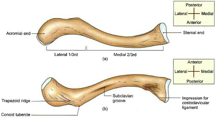
General features of right clavicle (a) Superior aspect (b) Inferior aspect


Features

It has 2 ends (Medial or Sternal, Lateral or Acromial end) and a shaft.

Lateral end 


  • It is flattend from above downwards.
  • It articulate with the acromian process of scapula to form acromioclavicular joint.

Medial end


  • It articulate with the sternum ( Manubrium sterni ) to form a joint which is called Sternoclavicular joint.
  • It receives attachment of interaclavicular ligament.

Shaft

It is divided into two parts anatomically...
1. Lateral one-third of clavicle.
2. Medial two-third of clavicle.

Lateral one-third of clavicle


  • It is flattend from above downwards.
  • It has 2 surfaces ( Superior and Inferior ) and 2 borders ( Anterior and Posterior ).

Superior Surface


  • It is subcutaneous.

Inferior Surface


  • It has coroid tubercle and trapizoid ridge, which gives attachment to the coroid and trapizoid part of the coracoclavicular ligament.

Anterior Border


  • It is concave anteriorly.
  • It provide attachment to the deltoid muscle.

Posterior Surface


  • It is convex backwards.
  • It gives attachment to the trapizeus.

Medial two-third of clavicle


  • It is quadrilateral in shape.
  • It has 4 surfaces ( Anterior, Posterior, Superior and Inferior ).

Anterior Surface


  • It is convex forwards.
  • It receives attachment of clavicular head of pectoralis major.

Posterior Surface


  • It is concave backwards.
  • It related to the internal jugular vein, subclavian vein and brachiocephalic vein.
  • Sternohyoid muscles originate from here.

Superior Surface


  • The clavicular head of  sterno cleido mastoid originate from this surface.


Inferior Surface


  • Several ligament attach on this surface like costoclavicular ligament and clavipectoral fascia.
  • Nutrient foramen also present on this surface, nutrient artery of clavicle passes though it.
Muscles attachment of clavicle, Clavicle
Attachment of the right clavicle (a) Superior aspect (b) Inferior aspect


Ossification


  • It ossifies from 2 primary and one secondary center ( Primary center appear during 5-6th week of intrauterine life for for shaft. Secondary center appear in puberty).


Ossification of clavicle, Clavicle
Ossification 


Applied


  • Most common fracture in this bone in road accident.
  • Weight loss of a patient can be assist clavicle. 


  You should also know about

Scapula

Scapula

Type - 

Flat

Quantity -

2 in numbers

Location 


  • It is posterolateral aspect of the thorax, against 2-7th rib.

Side Determination


  • Glenoid cavity looks laterally.
  • Coracoid process is directed forwards anteriorly.
  • Spinous process always posteriorly.


Features

Surfaces 

1. Costal Surface
2. Dorsal Surface

Costal Surface


  • It is also called as subscapular fossa.
  • It is concave.

Scapula, costal surface of scapula
Costal surface of right scapula

Dorsal Surface

A spine divide it (Dorsal Surface) into two parts...
1. Supra Spinous fossa or Surface.
2. Infra Spinous fossa or Surface.

Scapula, Dorsal surface of scapula, Dorsal surface
Dorsal surface of right scapula


Borders

1. Superior Border.
2. Lateral Border.
3. Medial Border

Superior Border


  • It is thin and shortest  border.
  • Suprascapular notch present on it.

Lateral Border

It is the thickest border.

Medial Border

It is also called as vertebral border.

Angles

1. Superior Angle.
2. Inferior Angle.
3. Lateral Angle.

Superior Angle

It covered by trapizeus muscle.

Inferior Angle

It covered by latissimus dorsi.

Lateral Angle


  • It bears glenoid cavity.
  • It is also called as glenoid angle.
  • It is also called as head of scapula.

Process

1. Spinus Process.
2. Acromial Process.
3. Coracoid Process.

Spinus Process


  • It is also called as spine of scapula, which is divides dorsal surface into supra spinus fossa and infra spinus fossa.
  • It has two surfaces (Superior and Inferior).

Acromial Process


  • It projects forwards from lateral end of spine.
  • It over hence the glenoid cavity.

Coracoid Process


  • It is directed forwards and slightly lateraly.
  • It arise from upper part of glenoid cavity.
  • Pectoralis major insert on it.

Attachment

1. Subscapularis arise from the medial two-third of the subscapular fossa.

2. Supraspinatus arises from the medial two-third of the supraspinous fossa.

3. Infraspinatus arise from the medial two-third of the infraspinous fossa.

Costal aspect, muscle attachment, scapula
Costal aspect muscle attachment


4. Deltoid arise from the lower border of the crest of the spine.

5. Trapezius is inserted into the upper border of the crest of the spine.

6. Serratus anterior is inserted along the medial border of the costal surface.

7. Long head of the biceps brachii arises from the supraglenoid tubercle, and short head from the lateral part of the tip of the coracoid process.

8. Coracobrachialis arises from the medial part of the tip of the coracoid process.

9. Pectoralis minor insert on coracoid process.

Dorsal aspect, muscle attachment, scapula
Dorsal aspect muscle attachment


10. Long head of the triceps brachii arises from the infraglenoid tubercle.

11. Teres minor arises by two slips on the dorsal surface along the lateral border. Circumflex scapular artery lies between the two slips.

12. Teres major arises from the dorsal aspect of the lateral border.

13. Levator scapulae is inserted along the dorsal aspect of the medial border.

14. Rhomboid minor and rhomboid major inserted into the medial border.

Right scapula muscle attachment, superior aspect, scapula
Superior aspect of the right scapula


15. Interior belly of the omohyoid arises from the upper border near the scapular notch.

16. Glenoidal labrum attached to the margin of the glenoid cavity.

17. Capsule of the acromioclavicular joint attached margin of the facet on the medial aspect of the acromion.

18. Coracoacromial ligament is attached to the coracoid process and acromion process.

19. Coracohumeral ligament is attached to the root of the coracoid process.

20. Coracoclavicular ligament is attached to the coracoid process.

Ossification


  • One primary center appear during 8th week of intrauterine life.
  • Seven secondary center appear for scapula.
  • All center fused up to age of 20 years.
  • Scapula's fusion are membranous. 

Ossification of scapula, ossification, scapula
Ossification of scapula


Applied

  • Paralysis of the serratus anterior causes 'winging' of the scapular.

T.M.J. (Temporomendibular joint)


T.M.J. (Temporomendibular joint)

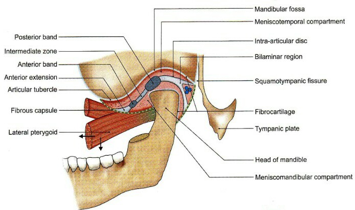
Type: condylar type of synovial joint.

Bones: Temporal and mandible.

Ligaments: Capsular ligament and temporomendibular ligament.

TMJ, temporomandibular joint, joint TMJ
Articular surface of the left temporomendibular joint


Movements: 

Protraction - pterigoid muscle

Retraction  - Digastric muscle, Massater muscle.

Dipration (Opening of mouth) - Lateral pterigoid muscle, infra hyoid muscle, digastric muscle.

Elevation of mandible - All muscles of the mastication.

Side to side movement of mandible -ipsilateral
pterigoid acting alternately.
Capsule of TMJ, Fibrous capsule and lateral ligament of the TMJ
Fibrous capsule and lateral ligament of the temporomandibular joint


Nerve supply: auriculo temporal nerve, Massetric nerve.

Applied: 
1. Dislocation.
2. Fracture.
3. Damage to auriculotemporal nerve.

Lungs Anatomy


Lungs

  • Lungs are essential organ of respiration.
  • Shape of lungs are like half-cone.
  • Lungs has an Apex & Base and 2 Surfaces and 3 Borders.
  • Two lungs are present in every human body.

Front of lungs, lungs with trachea
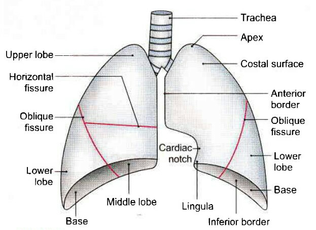
The trachea and lungs as seen from the front


Base - 

  • It is deeply concave and faces downwards.
  • It rest over the dom of diaphragm.

Apex - 

  • It is conical & directed upwards, towards the root of the neck.

Borders (3) -

Anterior Border

  • It is sharp and related to the anterior thoracic wall.
  • It's lower part has deep concavity of the left side which is called as cardiac notch.

Posterior Border

  • It is rounded & found the junction of costal and medial surface.
  • It corresponds with the head of the ribs.

Inferior Border

  • It is convex and sharp, it faces downwards.

Surfaces (2) -

Costal Surface

  • This surface is related to rib cage & sternum.

Medial Surface

It is divided into two parts...

1. Vertibral part - it is the posterior part of the medial surface and related to the vertibral bodies.

2. Mediastnal part - this surface is marked by hilum ( Hilum - it is depression from which structure enters or exit from here).
It is the large anterior part of the medial surface which is related to the mediastinum.

Side Determination -

  • Sharp anterior border faces forward.
  • Deep concave faces inferiorly.
  • Surface having hilum faces medially.

Right Lung - 

  • It has 2 fissures (Oblique, Transverse).
  • It has 3 lobes ( Superior, Middle and Inferior).

Mediastinal surface of right lung
Impression on the mediastinal surface of right lung


Left Lung -

  • It has only one fissure ( Oblique).
  • It has 2 lobes (Superior and Inferior).
  • The lower part of superior lobe is also called as lingula, which correspond with middle lobe of the right lung.

Mediastinal surface of left lung
Impression on the mediastinal surface of left lung


Difference between Right and Left Lung...

                   Right Lung                       Left Lung

1. Weight-  625gm                    -  565gm
2. Size -      Shorter & broader- longer & narrower
3. Fissure- Two                          -  one
4. Lobes -   Three                       - Two
5. Lingula - absent                    - present 
6. Cardiac notch - absent         - present 
7. BPS -      10                              - 8

Broncho pulmonary segments (BPS) 

  • It is difine as the developmental structural and functional unit of the lungs.
  • It consists of tersory bronchus and lungs tissue.


Right lung (10 - BPS)

  • Superior lobe has 3 BPS
  • Middle lobe has 2 BPS
  • Inferior lobe has 5 BPS

Left lung ( 8 - BPS)

  • Superior lobe has 3 BPS
  • Inferior lobe has 5 BPS

Bronchiopulmonary segments, BPS, segments of lungs
Bronchiopulmonary segments


Arterial Supply of Lungs

1. Pulmonary Arteries - one on each side ( Right and Left lungs).
2. Bronchial Arteries - one for right lung and two for left lung.

Venous drainage 

1. Pulmonary vein - two from right and two from left lungs (a superior and a inferior).
2. Bronchial vein - one from each lung.

Lymphatic drainage 

1. Pulmonary limph node.
2. Hilar limph node.
3. Trachiobronchial limph node.
4. Paratrachial limph node.

Nerve Supply 

  • Sympathetic - anterior and posterior pulmonary plexus.
  • Parasympathetic - vagus nerve.

Applied 

1. Pneumonectomy - removal of whole lung.
2. Lobectomy - removal of lobe.
3. Segmental resection - removal of BPS.


BRS Gross Anatomy by Kyung W. Chung, Harold M. Chung and Nancy L. Halliday

Meranazarya.blogspot.com

BRS Gross Anatomy by Kyung W. Chung, Harold M. Chung and Nancy L. Halliday


BRS Gross Anatomy book pdf

Download book pdf

About file:-

File type - pdf
File size - 57.05 MB
Pages - 525
Language - English 

Principles of Anatomy and Physiology by Geraro J. Tortora and Bryan Derrickson


Principles of Anatomy and Physiology by Geraro J. Tortora and Bryan Derrickson


Anatomy and physiology Tartora book pdf
Tartora book pdf


Download book pdf.

About file:-

File type - pdf
File size - 105.01 MB
Pages - 1281
Language - English 

Human Anatomy vol-3 ( Head-Neck and Brain) by BD Chourasia's

Meranazarya.blogspot.com

Human Anatomy vol-3 ( Head-Neck and Brain) by BD Chourasia's


Human anatomy BD Chourasia book pdf
BD chourasia Anatomy book pdf vol-3


Download book pdf

About file:-

File type - pdf
File size - 211.6 MB
Pages - 491
Language - English 

B D Chourasia's Human Anatomy Vol-2

B D Chourasia's Human Anatomy vol-2 ( Lower limb, Abdomen and pelvis) 6th edition


B D Chourasia's Human Anatomy Vol-2

CONTENTS....

SECTION : 1  LOWER LIMB

01 : Introduction (P-003)
02 : Bones of Lower Limb (P-006)
03 : Front of Thigh (P-040)
04 : Media Side of Thigh (P-059)
05 : Gluteal Region (P-065)
06 : Popliteal Fossa (P-076)
07 : Back of Thigh (P-083)
08 : Front, Lateral and Medial Sides of Leg and Dorsum of Foot (P-090)
09 : Back of Leg (P-103)
10 : Sole of Foot (P-113)
11 : Venous and Lymphatic Drainage Segmental Innervation, and Comparison of Lower and Upper Limbs (P-124)
12 : Joints of Lower Limb (P-136)
13 : Arches of Foot (P-159)
14 : Surface and Radiological Anatomy (P-165)
Appendix (P-172)

SECTION : 2  ABDOMEN AND PELVIS

15 : introduction and Osteology (P-183)
16 : Anterior Abdominal Wall (P-196)
17 : Male External Genital Organs (P-219)
18 : Abdominal Cavity and Peritoneum (P-229)
19 : Abdominal Part of Oesophagus and Stomach (P-248)
20 : Small and Large Intestines (P-257)
21 : Large Blood Vessels of the Gut (P-275)
22 : Extrahepatic Biliary Apparatus (P-287)
23 : Spleen, Pancreas and Liver (P-294)
24 : Kidney and Ureter (P-312)
25 : Suprarenal Gland and Chromaffin System (P-326)
26 : Diaphragm (P-331)
27 : Posterior Abdominal Wall (P-337)
28 : Perineum (P-350)
29 : Preliminary Consideration of Boundaries and Contents of Pelvis (P-367)
30 : Urinary Bladder and Urethra (P-372)
31 : Female Reproductive Organs (P-382)
32 : Male Reproductive Organs (P- 399)
33 : Rectum and /Anal Canal (P-407)
34 : Walls of Pelvis (P-419)
35 : Surface Marking of Abdomen and Pelvis (P-431)
36 : Radiological and Imaging Procedures (P-436)
Appendix 2 (P-441)


ABOUT BOOK

Name Book

B D Chourasia’s Human Anatomy (Vol. 2) 6th edition

Author/ Editior (6th edition)

B D Chourasia

Type

pdf.

Size

52.9 MB

Pages

910

Pages type

Colored

Quality (Good/average/bad)

Good


Similar Posts....

Endomyocardial Fibrosis