Liver
- Liver is the largest internal organ of the body.
- Liver is the largest exocrine gland of the body.
- Liver is the main heat producing organ of the body.
- 1500 ml/min blood pass-out from the liver.
- 400 ml blood always present in the liver.
- Chief cells of the liver are called hepatocyte which forms 80% mass of the liver.
- Synonym
- Location
- Measurement
- Presenting parts
- Relation
- Ligaments
- Porta hepatis
- Hepatic segments
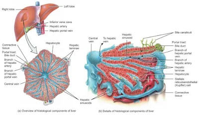
- Histology
- Functions
- Blood supply
- Lymphatic drainage
- Nerve supply
- Applied anatomy
 |
| Front view of the liver |
Synonym
Haper, hepta, biochemical lab, kitchen of the body.
Location
Measurements
Weight - 1200 - 1500gms ( male - 1400-1600, female - 1200 - 1400gms)
Length - 26cm in male and 25cm in female average
Thickness - 22cm in adult male and 21cm in adult female average
Height - 7cm average
Color - Reddish brown
Consistency - soft
Quantity - one in number
Shape - wedge shape
Thickness - 22cm in adult male and 21cm in adult female average
Height - 7cm average
Color - Reddish brown
Consistency - soft
Quantity - one in number
Shape - wedge shape
Presenting parts
Surfaces ( 5 ) - Superior, Inferior, Anterior, Posterior, Right.
Border - Inferior
Lobes -
a. Necessary lobes - Right lobe and left lobe.
b. Accessory lobes - Caudate and quadrate lobe.
Surfaces
Superior surface
It is quadrilateral and shows a concavity in the middle called cardiac impression
Inferior surface
It is quadrilateral and is directed downwards and down to the left. Gastric impression, fissure for ligamentum teres, quadrate lobe, fossa for gallbladder and colic impression on it,
Anterior surface
It is triangular and slightly convex. Falciform ligament is attached on it.
Posterior surface
It is triangular and middle part of this surface shows a concavity for vertebral column. Bare area, groove for inferior vena cava, caudate lobe, fissure for ligamentum venosum and oesophageal impression.
Right surface
It is quadrilateral and convex. it is related to the diaphragm opposite to the 7th to 11th ribs in the mid axillary line.
Border
Inferior border
It is sharp anteriorly and interlobar notch for ligamentum teres and a cystic notch for the fundus for gallbladder present on it.
Lobes
Liver is divided into two lobes (right and left ) by the falciform ligament anteriorly and superiorly, fissure for ligamentum teres inferiorly and fissure for ligamentum venosum posteriorly.
Right lobe
It is much larger than the left lobe and forms 5/6 part of the liver. caudate (posterior surface) and quadrate (inferior surface ) lobe present on it.
Caudate lobe
it is situated on the posterior surface.
Quadrate lobe
It is situated on the inferior surface.
Left lobe
It is the much smaller than the right lobe and forms 1/6 part of the liver. tuber omentale or omental tuberosity present on it.
Relation
a. Peritoneal relation
Most of the liver is covered by the peritoneum except triangular bare area, groove for inferior vena cava, fossa for gallbladder, area attachment for lesser omentum and ligamentum venosum.
b. Visceral realation
Anteriorly
Xiphoid process, anterior abdominal wall, diaphragm and falciform ligament,
Posteriorly
Inferior vena cava, ligamentum venosum, abdominal part of oesophagus, bare area and caudate lobe.
Superiorly
Diaphragm
Inferiorly
Stomach, ligamentum teres and quadrate lobe.
 |
| Relations of Posterior and inferior surface of the liver |
Right
Diaphragm and mid axillary line.
Ligaments
a. True ligaments
I. Ligamentum venosum (posteriorly) - remnant part of the ductus venosum.
II. Ligamentum teres (inferiorly) - remnant part of the umbilical cord. It is also called round ligament of the liver.
b. False ligaments
These are peritoneal folds.
These are 5 in number...
I. Falciform ligament
II. Coronary ligament
III. Right triangular ligament
IV. Left triangular ligament
V. Lessor omentum
Porta heapatis
It is also called as gateway of liver.
It situated on the posteroinferior surface of right lobe of the liver.
It's length about 5cm.
Portal vein and hepatic artery passes through it.
Hepatic segments
On the basis of intrahepatic distribution of the hepatic artery, portal vein and biliary duct, liver can be divided right and left functional lobes. These lobe don't correspond to the anatomical lobes.
Right anatomical lobe is sub divided into these hepatic segments...
- Right anterior ( V and VIII )
- Right posterior ( VI and VII )
Left anatomical lobe are sub divided into these hepatic segments...
- Left lateral ( II and III )
- Left medial ( I and IV )
 |
| Segments of the liver |
Histology
 |
| Histology of liver (a). Portal lobule, (b). Liver acinus |
Functions
- Metabolism - carbohydrates, fats and proteins.
- Synthesis - bile and prothrombin.
- Excretion - drugs, toxins, poisons, cholesterol, bile pigments, and heavy metals.
- Protective - conjugation, destruction, phagocytosis, antibody formation and excretion.
- Storage - glycogen, fat, vitamins ( A,D,E,K and B12 ) and minerals ( iron ).
Blood supply
Arterial supply -
- Hepatic artery - 20% blood supply for it.
- Portal vein - 80% blood supply for it.
Venous drainage -
Lymphatic drainage
The liver produce a large amount of lymph which is estimated 25 - 50% of total lymph.
- Hepatic lymph node
- Paracardial lymph node
- coeliac lymph node
- Thoracic duct
- Caval
Nerve supply
Hepatic nervous plexus which contain sympathetic and parasympathetic nerve fibers.
- Sympathetic fibers - celiac plexus
- Parasympathetic fibers - vagal fibers.
Clinical Anatomy of the Liver
- Location and Examination: The liver is located in the infrasternal angle and can be examined through percussion, although it is usually not palpable due to the tone of the recti muscles and its softness.
- Position: In the median plane, the lower border of the liver lies on the transpyloric plane, about a hand’s breadth below the xiphisternal joint. In women and children, this border often projects slightly below the right costal margin.
- Liver Enlargement: The liver may enlarge towards the right iliac fossa. The spleen can also enlarge in the same direction.
- Inflammation (Hepatitis): Liver inflammation, known as hepatitis, can be infective or amoebic.
- Cirrhosis: In cases of malnutrition, liver tissue may undergo fibrosis and shrink, leading to cirrhosis.
- Liver Biopsy: A liver biopsy is performed by inserting a needle through the right 9th intercostal space, passing through both the pleural and peritoneal cavities.
- Metastasis: The liver is a common site for metastatic tumors, as venous blood from the gastrointestinal tract drains into the liver via the portal vein.
- Blood Supply: The liver receives blood from both the hepatic artery and the portal vein, which lie in the free margin of the lesser omentum. Bleeding can be controlled by compressing this area (Pringle’s maneuver).
- Liver Resection and Regeneration: Up to 80% of the liver can be safely removed during surgery, with the liver capable of regenerating within 6-12 months.
- Liver Transplantation: Liver transplants involve grafting an organ from a donor, with various anastomoses of the veins, arteries, and bile ducts. A partial liver from a healthy donor can also be transplanted.
- Portal Hypertension: In severe cases, procedures like TIPS (transjugular intraparenchymal portosystemic shunt) are used to reduce portal hypertension.
- Cirrhosis Symptoms: Advanced cirrhosis can lead to "caput medusae," where distended veins are visible around the umbilicus.
Images are copied from BD chourasia and Tortora books


No comments:
Post a Comment
Please do not enter any spam link in the comment box