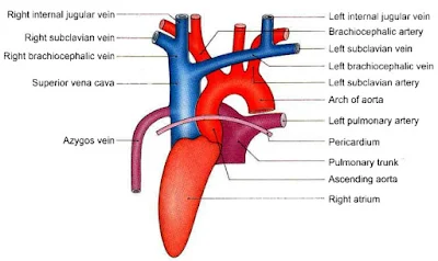
Superior vena cava

Superior vena cava

  • Superior vena cava is a large venous channel which collects blood from the upper half of the body and drain into the right atrium.
  • Superior vena cava is about 7cm long.
  • It is formed by the union of the right and left brachiocephalic or innominate veins.
  • It begins behind the lower border of the first right costal cartilage close to the sternum.
  • It terminates by the opening into the upper part of the right atrium behind the third right costal cartilage.
  • It has no valves.
Superior vena cava, vena cava, Aas, alfaaz-e-aas
Superior vena cava and it's relations


Relations

Anterior

  • Chest wall.
  • Internal thoracic vessels.
  • Anterior margin of the right lung and pleura.
  • The vessel is covered by pericardium in its lower half.

Posterior

  • Trachea
  • Right vagus
  • Root of the right lung

Medial

  • Ascending aorta
  • Brachiocephalic artery

Lateral

  • Right phrenic nerve and vessels
  • Right pleura and lung

Tributaries of the superior vena cava

  1. Azygos vein ( at the level of second costal cartilage ).
  2. Several small mediastinal and pericardial veins drain into the vena cava.

Applied anatomy

  1. Obstruction of superior vena cava above the opening of the azygos vein.
  2. Obstruction of the superior vena cava below the opening of the azygos vein.
  3. Obstruction of the superior vena cava is indicates the mediastinal syndrome.


Image copied from BD Chourasia book
Comments

Popular posts from this blog

Histology text & Atlas by Brijesh kumar

Histology text & Atlas by Brijesh kumar Download book pdf About file:- File type - pdf File size - 26.35 MB Pages - 392 Language - English 

Practical Medicine (P. J. Mehta's)

Practical Medicine (P. J. Mehta's) 20th edition CONTENTS PART-1 CLINICAL CASES 1. History Taking and Symptomatology 2. General Examination 3. Abdomen 4. Respiratory System 5. Cardiovascular System 6. Central Nervous System PART-2 TABLE-WORK 7. Medical Emergencies 8. Electrocardiography 9. Radiology 10. Instruments 11. Procedures 12. Hematology 13. Clinical Pathology 14. Pathology Specimens 15. Drugs ABOUT BOOK Name Book Practical Medicine (P. J. Mehta’s) Author/ Editior (20 th edition) SP Mehta, SR Joshi, Nihar P Mehta / Dr. Hardik shah, Dr. Sunita Iyer, Dr. Nikesh Jain, Dr. Rajiv Shah. Type pdf. Size 69.47 MB Pages 630 Pages type Colored Quality (Good/average/bad) Good D ownload Now Similar Posts :- 1.  Park’s Textbook of PSM 2.  Review of Preventive and Social Medicine (Vivek Jain) 3.  Medicine (Exam Preparatory

Lysosomes

Name :- Lysosomes, garbage system of the cell, suicidal bag of the cell Properties : These are membrane bound vesicular structures founds throughout the cytoplasm. These are formed by the process of packaging in the golgi apparatus. Enzymes of lysosomes are synthesized in rough endoplasmic reticulum. Among the organelles, the lysosomes have the thickest covering membrane that is formed by a bilayered lipid material . Lysosomes have 50 different hydrolytic enzymes, known as acid hydroxylases. Types of Lysosomes : Primary lysosomes, this is an inactive of form of lysosomes. Secondary lysosomes, this is an active form of lysosomes (lysosomes active during phagosome or endosome and at this time lysosomes pH become acidic and enzymes are activate). Lysosomal Enzymes :   Proteases :- which hydrolyze the proteins into amino acids. Lipases :- which hydrolyze the lipids into fatty acids and glycerides. Amylases :- which hydrolyze the polysaccharides into glucose. Nucleases :- whi