Hip bone
Names: innominate, irregular, Hip bone.
Bones: ilium ( latin- loin) superiorly , ischium (greek- hip joint) posteroinferiorly, pubis ( latin- genital area) anteroinferiorly.
Joint formation:
Hip joint - joint between head of femur and acetabulum.
Pubic symphysis - between two hip bone anteriorly.
Site determination:
1. The acetabulum is directed laterally.
2. Ilium situated superiorly.
3. The obturator foramen lies below the acetabulum.
Anatomical position:
1. The pubic tubercle and anterior superior iliac spine lie in the same coronal plane.
2. The pelvic surface of the body of the pubis is directed backwards and upwards.
3. Symphyseal surface lies medialy.
 |
| Hip bone |
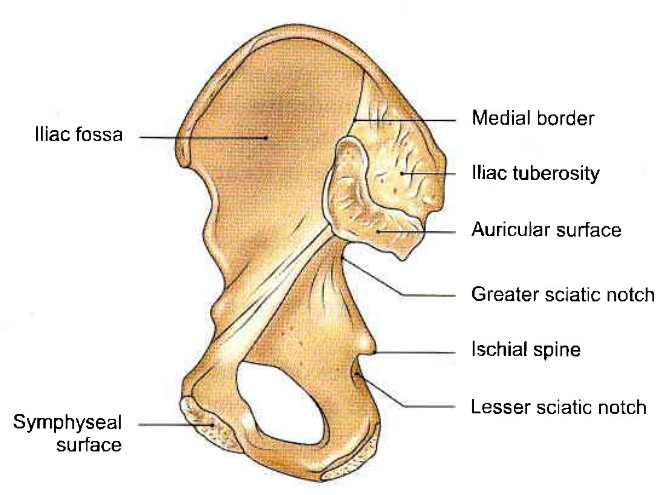
Inner surface of right hip bone
 |
| Hip bone |
Outer surface of right hip bone
Ilium
Ilium or flank form upper expanded plate like part of hip bone. It forms upper two fifth(2/5) of the acetabulum.
It has...
Three borders: anterior, posterior, medial.
Three surface: gluteal surface, iliac surface or iliac fossa, sacropelvic surface.
One upper end called iliac crest.
One lower end which is smaller and is fused with the pubis and the ischium at the acetabulum.
Iliac crest:
It is a broad convex ridge forming upper end of the ilium.
The highest point of the iliac crest - it is situated a little behind the mid point of the crest. It lies at the level of interval between the spines of vertebrae L3- L4.
Borders
Borders
Anterior border - two prominent parts present here... a. Anterior superior iliac spine.
b. Anterior inferior iliac spine.
Posterior border - it contain two prominent parts and a notch...
a. posterior superior iliac spine.
b. Posterior inferior iliac spine.
c. Greater sciatic notch.
Medial border - it extend inner or pelvic surface of the ilium from the iliac crest to the iliopubic eminence. It separate the iliac fossa from the sacropelvic surface. Its lower part rounded forms the iliac part of the arcuate line or inlet of pelvis.
Surfaces
Gluteal surface - it is the outer surface of the ilium, which is convex in front and concave behind like the iliac crest.
It is divided into four areas by three gluteal line...
a. Posterior gluteal line.
b. Anterior gluteal line.
c. Inferior gluteal line.
Iliac surface - it is the large concave area on the inner surface of the ilium, situated in front of its medial border. It forms the lateral wall of false pelvis.
Sacropelvic surface - it is the uneven area on the inner surface of the ilium, situated bhind the its medial border.
It is subdivided into three parts...
a. Iliac tuberosity - it is the upper larged roughened area.
b. Auricular surface - it is articular but pitted.
c. Pelvic surface - it is smooth and lies anteroinferior to the auricular surface. It forms a part of lateral wall of true pelvis. Along the upper border of greater sciatic notch, this surface is marked by the preauricular sulcus. This sulcus is deeper in female than in male.
Attachment of the ilium
1. Anterior superior iliac spine:
a. Lateral end of inguinal ligament.
b. It gives origin sartorius muscle, the origin extends onto the upper half of the notch below the supine.
2. Outer lip of iliac crest:
a. Fascia lata attach here.
b. Origin to the tensor fascia lata in front of the tubercle.
c. Insertion to the external oblique muscle in its anterior two-third.
d. Origin to latissimus dorsi just behind highest point of the crest.
e. Iliotibial tract attach here.
3. Intermediate area of iliac crest:
a. Origin internal oblique muscle from its anterior two-third.
4. Inner lip of the iliac crest:
a. Origin to the transversus abdominis from its anterior two-third.
b. Fascia iliaca and fascia transversalis attach on lts anterior two-third.
c. Origin to the quadratus lumborum from its posterior one-third.
d. Attachment of the thoracolumbar fascia around the attachment of the quadratus lumborum.
5. Dorsl segment of iliac crest:
a. Origin to the gluteus maximus from its lateral slope.
b. Origin to the erector spinae from its medial slope.
c. Interosseous and sacroiliac ligaments are attach to the medial margin deep to the attachment of the erector spinae.
6. Upper half of the anterior superior iliac spine:
a. Origin to the straight head of the rectus femoris.
b. Its lower part gives attachment to the iliofemoral ligament.
7. Posterior border of the ilium:
a. Upper fibres of the sacrotuberous ligament attach above the greater sciatic notch.
b. Few fibres of piriformis origin from upper margin of the greater sciatic notch.
8. Gluteal surface:
a. Upper fibres of gluteus maximus origin from behind the gluteal line.
b. Gluteus medius arise from the area between the anterior and posterior gluteal line.
c. Gluteus minimus arise from the area between the anterior and inferior gluteal line.
d. The reflected head of the rectus femoris arise from the groove above the acetabulum.
e. The capsular ligament of the hip joint is attached along the margin of acetabulum.
9. Iliac fossa:
a. Origin to the iliacus from it's upper two third.
b. Lower groove is covered by the iliac bursa.
10. Iliac tuberosity:
a. Interosseous sacroiliac ligament in its greater part.
b. The dorsal sacroiliac ligament posteriorly.
c. Iliolumbar ligament superiorly.
11. Convex margin of the auricular surface:
a. It gives attachment to scroiliac ligaments.
12. Pelvic surface:
a. Lower fibre of the ventral sacroiliac ligament from preauricular sulcus.
b. Few fibres of the piriformis origin from lateral to the preauricular sulcus.
c. Upper half of the obturator internus origin from the pelvic surface.
Surfaces
Gluteal surface - it is the outer surface of the ilium, which is convex in front and concave behind like the iliac crest.
It is divided into four areas by three gluteal line...
a. Posterior gluteal line.
b. Anterior gluteal line.
c. Inferior gluteal line.
Iliac surface - it is the large concave area on the inner surface of the ilium, situated in front of its medial border. It forms the lateral wall of false pelvis.
Sacropelvic surface - it is the uneven area on the inner surface of the ilium, situated bhind the its medial border.
It is subdivided into three parts...
a. Iliac tuberosity - it is the upper larged roughened area.
b. Auricular surface - it is articular but pitted.
c. Pelvic surface - it is smooth and lies anteroinferior to the auricular surface. It forms a part of lateral wall of true pelvis. Along the upper border of greater sciatic notch, this surface is marked by the preauricular sulcus. This sulcus is deeper in female than in male.
Attachment of the ilium
 |
| Hip bone iliac crest |
1. Anterior superior iliac spine:
a. Lateral end of inguinal ligament.
b. It gives origin sartorius muscle, the origin extends onto the upper half of the notch below the supine.
2. Outer lip of iliac crest:
a. Fascia lata attach here.
b. Origin to the tensor fascia lata in front of the tubercle.
c. Insertion to the external oblique muscle in its anterior two-third.
d. Origin to latissimus dorsi just behind highest point of the crest.
e. Iliotibial tract attach here.
3. Intermediate area of iliac crest:
a. Origin internal oblique muscle from its anterior two-third.
4. Inner lip of the iliac crest:
a. Origin to the transversus abdominis from its anterior two-third.
b. Fascia iliaca and fascia transversalis attach on lts anterior two-third.
c. Origin to the quadratus lumborum from its posterior one-third.
d. Attachment of the thoracolumbar fascia around the attachment of the quadratus lumborum.
5. Dorsl segment of iliac crest:
a. Origin to the gluteus maximus from its lateral slope.
b. Origin to the erector spinae from its medial slope.
c. Interosseous and sacroiliac ligaments are attach to the medial margin deep to the attachment of the erector spinae.
6. Upper half of the anterior superior iliac spine:
a. Origin to the straight head of the rectus femoris.
b. Its lower part gives attachment to the iliofemoral ligament.
7. Posterior border of the ilium:
a. Upper fibres of the sacrotuberous ligament attach above the greater sciatic notch.
b. Few fibres of piriformis origin from upper margin of the greater sciatic notch.
8. Gluteal surface:
a. Upper fibres of gluteus maximus origin from behind the gluteal line.
b. Gluteus medius arise from the area between the anterior and posterior gluteal line.
c. Gluteus minimus arise from the area between the anterior and inferior gluteal line.
d. The reflected head of the rectus femoris arise from the groove above the acetabulum.
e. The capsular ligament of the hip joint is attached along the margin of acetabulum.
9. Iliac fossa:
a. Origin to the iliacus from it's upper two third.
b. Lower groove is covered by the iliac bursa.
10. Iliac tuberosity:
a. Interosseous sacroiliac ligament in its greater part.
b. The dorsal sacroiliac ligament posteriorly.
c. Iliolumbar ligament superiorly.
11. Convex margin of the auricular surface:
a. It gives attachment to scroiliac ligaments.
12. Pelvic surface:
a. Lower fibre of the ventral sacroiliac ligament from preauricular sulcus.
b. Few fibres of the piriformis origin from lateral to the preauricular sulcus.
c. Upper half of the obturator internus origin from the pelvic surface.
Pubis
It forms the anteroinferior part of the hip bone and the anterior One- fifth of the acetabulum , forms the anterior boundary of the obturator foramen.
It has...
A body superiorly.
A superior ramus superolaterally.
A inferior ramus inferolterally.
Body of pubis:
This is flattened from before backward, and has...
1. A superior border called pubic crest.
2. A pubic tubercle at the lateral end of the pubic crest.
3. Three surfaces anterior, posterior and medial.
Anterior surface - it is directed downward, forwards and slightly laterally. it is rough superomedially and smooth elsewhere.
Posterior or pelvic surface - it is smooth. it is directed upwards and backwards. it forms the anterior wall of the true pelvis and is related to the urinary bladder.
Medial or symphyseal surface - it articulates with the opposite pubis to form the pubic symphysis.
Superior ramus :
It extends from the body of the pubis to the acetabulum above the obturator foramen.
It has...
a. Three borders.
b. Three surfaces.
Borders:
a. Superior border or pectineal line or pecten pubis.
b. Anterior border or obturator crest.
c. Inferior border.
Surfaces:
a. Pectineal surface.
b. Pelvic surface.
c. Obturator surface.
Inferior ramus:
it extends from the body of the pubis to the ramus of the ischium to form the conjoined ischiopubic rami.
Attachment and relations of the pubis:
1. Pubic tubercle - in males the tubercle is crossed by the spermatic cord.
a. Medial end of the inguinal ligament.
b. Ascending loops of the cremaster muscle.
2. The medial part of the pubic crest is crossed by the medial head of the rectus abdominis.
Lateral head of the rectus abdominis and pyramedialis origin from lateral part of the crest.
3. Anterior surface of the body:
a. Anterior pubic ligament medially.
b. Origin adductor longus in the angle between crest and symphysis.
c. Origin to the gracilis and form the inferior ramus.
d. Origin to the adductor brevis lateral to the origin of the gracilis.
e. Origin to the obturator externus near the margin of the operator foramen.
4. Posterior surface of the body:
a. Origin to the levator ani from its middle part.
b. Origin to the obturator internus laterally.
c. Attachment to the puboprostatic / pubovesical ligament medial to the attachment of the levator ani.
5. Pectineal line:
a. The conjoint tendon at the medial end.
b. The lacunar ligament at the medial end, in front of the attachment of the conjoint tendon.
c. Pectineal ligament of cooper.
d. Pectineus muscle arise from here.
e. The fascia covering of the pectineus.
f. The psoas minor insert here.
6. Pectineal surface:
a. Pectineus from upper part.
7. Pelvic surface is crossed by round ligament of the uterus in females and by the ductus deference in males.
8. The obturator groove transmits the obturator vessels and nerve.
Ischium
The ischium forms the posteroinferior part of the hip bone and the adjoining two fifth of the acetabulum. It forms the posterior boundary of the oturator foramen.
It has...
a. A body.
b. A ramus.
Body of the ischium:
This is a thick and massive mass of bone that lies below and behind the acetabulum.
It has...
a. Two ends - upper and lower.
b. Three borders - anterior, posterior and lateral.
c. Three surfaces - femoral, dorsal and pelvic.
Ends
1. Upper end - it forms the posteriinferior two- fifth of the acetabulum.
2. Lower end - it forms the ischial tuberosity. It gives off the ramus of the ischium which forms an acute angle with the body.
Borders
1. Anterior border - it forms the posterior margin of the obturator foramen.
2. Posterior border - it is continuous above with the posterior border of the ilium. It also form part of the greater sciatic notch. Below the notch posterior margin shows a projection called the ischial spine. below the spine the posterior border show a concavity is called the lesser sciatic notch.
3. Lateral border - it forms the lateral margin of the ischial tuberosity except the upper end where it is rounded.
Surfaces
1. Femoral surface - it lies between the anterior and lateral borders.
2. Dorsal surface - it continuous above with the gluteal surface of the ilium.
3. Pelvic surface - it is smooth and forms part of the lateral wall of the true pelvis.
Ramus
The inferior ramus of the pubis unites with the ramus of the ischium on the medial side of the obturator foramen.
It has...
a. Two borders.
b. Two surfaces.
Borders
1. Upper border - it forms part of the margin of the obturator foramen.
2. Lower border - it forms the pubic arch along with the corresponding border of the bone of the opposite side.
Surfaces
1. Inner surface - it is convex and smooth. It is divided into three areas upper, middle and lower by two ridges.
2. Outer surface - it is rough for the attachment of the muscles.
Attachment and relations of the ischium
1. Ischial spine:
a. Attach sacrospinous ligament.
b. Origin for the posterior fibres of the levator ani.
2. Lesser sciatic notch:
a. Origin to the superior and inferior gemelli.
3. Femoral surface:
a. Obturator externus along the margin of the obturator foramen.
b. Quadratus femoris along the lateral border of the upper part of the ischial tuberosity.
4. Dorsal surface:
a. Piriformis, the sciatic nerve and nerve of the quadratus femoris.
5. Ischial tuberosity:
a. Origin semimembranosus from superolateral area.
b. Semitendinosus and the long head of the biceps femoris from inferomedial area.
c. Adductor magnus from outer lower area.
d. Sacrotuberous ligament.
e. Ischiofemoral ligament.
6. Greater part of the pelvic surface:
a. Origin obturator internus.
7. Conjoined ischiopubic rami:
a. Obturator membrane attach on upper part.
b. Fascia lata and membranous layer of superficial fascia or colles'fascia of the perineum attach with lower border.
c. Origin obturator externus from near the obturator margin of both rami.
d. Origin adductor brevis and gracilis from pubic ramus.
e. Origin adductor magnus from the ischial ramus.
f. Perineal membrane is attach to the inner surface lower ridge.
g. Inner surface upper area gives origin to the obturator internus.
h. Inner surface middle area gives origin transvers perinei.
i. Lower area gives attachment to crus penis and gives origin ischiocavernosus and to the superficial transversus perinei.
Acetabulum
1. it is a deep Cup shaped hemispherical cavity on the lateral aspect of the hip bone, about its centre.
2. It is directed laterally, downwards and forwards.
3. It has a margin deficient inferiorly, it is also called acetabular notch.
4. Non articular roughned floor is called acetabular fossa.
5. horseshoe shaped articular surface or lunate surface is seen on the anterior, superior, and posterior part of the acetabulum. it is line with high lined cartilage, and articulates with the head of the femur to form the hip joint. the fibrocartilaginous acetabular labrum is attached to the margin of the acetabulum, is deepens the acetabular cavity.
Obturator foramen
1. This is a large gap in the hip bone, situated anteroinferior to the acetabulum, between the pubis and the ischium.
2. It is large and oval in males, and small and triangular in female.
Ossification
It 's ossification is cartilaginous from three primary and five secondary centre. Complete bone ossified till 20-25 year of age.
Applied
1. Iliac crest used for taking bone marrow biopsy in cases of anemia or leukemia.
2. Weaver's bottom -- person sitting for a long period of time may get inflammation of their ischial tuberosity.
This is flattened from before backward, and has...
1. A superior border called pubic crest.
2. A pubic tubercle at the lateral end of the pubic crest.
3. Three surfaces anterior, posterior and medial.
Anterior surface - it is directed downward, forwards and slightly laterally. it is rough superomedially and smooth elsewhere.
Posterior or pelvic surface - it is smooth. it is directed upwards and backwards. it forms the anterior wall of the true pelvis and is related to the urinary bladder.
Medial or symphyseal surface - it articulates with the opposite pubis to form the pubic symphysis.
Superior ramus :
It extends from the body of the pubis to the acetabulum above the obturator foramen.
It has...
a. Three borders.
b. Three surfaces.
Borders:
a. Superior border or pectineal line or pecten pubis.
b. Anterior border or obturator crest.
c. Inferior border.
Surfaces:
a. Pectineal surface.
b. Pelvic surface.
c. Obturator surface.
Inferior ramus:
it extends from the body of the pubis to the ramus of the ischium to form the conjoined ischiopubic rami.
Attachment and relations of the pubis:
1. Pubic tubercle - in males the tubercle is crossed by the spermatic cord.
a. Medial end of the inguinal ligament.
b. Ascending loops of the cremaster muscle.
2. The medial part of the pubic crest is crossed by the medial head of the rectus abdominis.
Lateral head of the rectus abdominis and pyramedialis origin from lateral part of the crest.
3. Anterior surface of the body:
a. Anterior pubic ligament medially.
b. Origin adductor longus in the angle between crest and symphysis.
c. Origin to the gracilis and form the inferior ramus.
d. Origin to the adductor brevis lateral to the origin of the gracilis.
e. Origin to the obturator externus near the margin of the operator foramen.
4. Posterior surface of the body:
a. Origin to the levator ani from its middle part.
b. Origin to the obturator internus laterally.
c. Attachment to the puboprostatic / pubovesical ligament medial to the attachment of the levator ani.
5. Pectineal line:
a. The conjoint tendon at the medial end.
b. The lacunar ligament at the medial end, in front of the attachment of the conjoint tendon.
c. Pectineal ligament of cooper.
d. Pectineus muscle arise from here.
e. The fascia covering of the pectineus.
f. The psoas minor insert here.
6. Pectineal surface:
a. Pectineus from upper part.
7. Pelvic surface is crossed by round ligament of the uterus in females and by the ductus deference in males.
8. The obturator groove transmits the obturator vessels and nerve.
Ischium
The ischium forms the posteroinferior part of the hip bone and the adjoining two fifth of the acetabulum. It forms the posterior boundary of the oturator foramen.
It has...
a. A body.
b. A ramus.
Body of the ischium:
This is a thick and massive mass of bone that lies below and behind the acetabulum.
It has...
a. Two ends - upper and lower.
b. Three borders - anterior, posterior and lateral.
c. Three surfaces - femoral, dorsal and pelvic.
Ends
1. Upper end - it forms the posteriinferior two- fifth of the acetabulum.
2. Lower end - it forms the ischial tuberosity. It gives off the ramus of the ischium which forms an acute angle with the body.
Borders
1. Anterior border - it forms the posterior margin of the obturator foramen.
2. Posterior border - it is continuous above with the posterior border of the ilium. It also form part of the greater sciatic notch. Below the notch posterior margin shows a projection called the ischial spine. below the spine the posterior border show a concavity is called the lesser sciatic notch.
3. Lateral border - it forms the lateral margin of the ischial tuberosity except the upper end where it is rounded.
Surfaces
1. Femoral surface - it lies between the anterior and lateral borders.
2. Dorsal surface - it continuous above with the gluteal surface of the ilium.
3. Pelvic surface - it is smooth and forms part of the lateral wall of the true pelvis.
Ramus
The inferior ramus of the pubis unites with the ramus of the ischium on the medial side of the obturator foramen.
It has...
a. Two borders.
b. Two surfaces.
Borders
1. Upper border - it forms part of the margin of the obturator foramen.
2. Lower border - it forms the pubic arch along with the corresponding border of the bone of the opposite side.
Surfaces
1. Inner surface - it is convex and smooth. It is divided into three areas upper, middle and lower by two ridges.
2. Outer surface - it is rough for the attachment of the muscles.
Attachment and relations of the ischium
1. Ischial spine:
a. Attach sacrospinous ligament.
b. Origin for the posterior fibres of the levator ani.
2. Lesser sciatic notch:
a. Origin to the superior and inferior gemelli.
3. Femoral surface:
a. Obturator externus along the margin of the obturator foramen.
b. Quadratus femoris along the lateral border of the upper part of the ischial tuberosity.
4. Dorsal surface:
a. Piriformis, the sciatic nerve and nerve of the quadratus femoris.
5. Ischial tuberosity:
a. Origin semimembranosus from superolateral area.
b. Semitendinosus and the long head of the biceps femoris from inferomedial area.
c. Adductor magnus from outer lower area.
d. Sacrotuberous ligament.
e. Ischiofemoral ligament.
6. Greater part of the pelvic surface:
a. Origin obturator internus.
7. Conjoined ischiopubic rami:
a. Obturator membrane attach on upper part.
b. Fascia lata and membranous layer of superficial fascia or colles'fascia of the perineum attach with lower border.
c. Origin obturator externus from near the obturator margin of both rami.
d. Origin adductor brevis and gracilis from pubic ramus.
e. Origin adductor magnus from the ischial ramus.
f. Perineal membrane is attach to the inner surface lower ridge.
g. Inner surface upper area gives origin to the obturator internus.
h. Inner surface middle area gives origin transvers perinei.
i. Lower area gives attachment to crus penis and gives origin ischiocavernosus and to the superficial transversus perinei.
 |
| Hip bone Muscles attachment |
 |
| Hip bone Muscles attachment |
Acetabulum
1. it is a deep Cup shaped hemispherical cavity on the lateral aspect of the hip bone, about its centre.
2. It is directed laterally, downwards and forwards.
3. It has a margin deficient inferiorly, it is also called acetabular notch.
4. Non articular roughned floor is called acetabular fossa.
5. horseshoe shaped articular surface or lunate surface is seen on the anterior, superior, and posterior part of the acetabulum. it is line with high lined cartilage, and articulates with the head of the femur to form the hip joint. the fibrocartilaginous acetabular labrum is attached to the margin of the acetabulum, is deepens the acetabular cavity.
Obturator foramen
1. This is a large gap in the hip bone, situated anteroinferior to the acetabulum, between the pubis and the ischium.
2. It is large and oval in males, and small and triangular in female.
Ossification
It 's ossification is cartilaginous from three primary and five secondary centre. Complete bone ossified till 20-25 year of age.
Applied
1. Iliac crest used for taking bone marrow biopsy in cases of anemia or leukemia.
2. Weaver's bottom -- person sitting for a long period of time may get inflammation of their ischial tuberosity.


No comments:
Post a Comment
Please do not enter any spam link in the comment box