Infarction
Definition –
A necrotic area is called infarct and the process of infarct formation is
called infarction.
Causes –
Ischaemic necrosis (most common), stagnant hypoxia (venous obstruction),
thrombosis, embolism, narrowing of the coronary artery (atherosclerosis) etc.
Types of
infarcts –
1. According to
colour …
a. Pale or anemic infarct – It is
due to arterial occlusion, commonly seen in compact organ like heart, kidney
and spleen.
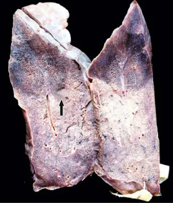
b. Red or haemorrhagic infarct – It
is due to obstruction of the artery, commonly seen in soft loose tissue e.g.
lungs and intestine.
 |
| Haemorrhagic Infarct of the Lung |
2. According to
the age … Recent and old.
3. According to
the presence or absence of the infection…
a. Bland – Free from bacterial
contamination
b. Infected – Infected with
bacterial contamination.
4. According to
the type of action…
a. Bacteriostatic – It inhibit growth
of bacteria e.g. tetracycline, erythromycin, sulfonamide etc.
b. Bactericidal – It kills bacteria
e.g. refampicin, penicillin etc.
5. According to
the spectrum of activity… narrow and broad spectrum.
PATHOGENESIS –
- Localized hyperaemia or congestion may occur due to obstruction of blood supply.
- Within few hours edema and haemorrhage may occur.
- Cellular change such as cloudy swelling appear early, necrosis occur within 12-15 hours.
- Progressive proteolysis of necrotic tissue and there is lysis of red cells.
- Acute inflammatory reactions appear in surrounding tissue.
- Blood pigments like haematoidin and haemosiderin deposit in the infarct.
- Following this granulation tissue starts to appears at the margin, finally infarct is replaced by collagen fibers and a scar is formed.
GROSSLY – Infarcts of solid arms are usually wedge shaped with apex at the obstructed artery and base of the surface of the organ. Most infarcts become pale due to breakdown of red cells but pulmonary infarcts never become pale due to extensive blood supply.
Cerebral infarcts appear with central softening due to liquefaction
necrosis (encephalomalacia), recent infarcts are generally slightly elevated
over the surface but old infarcts are shrunken and depressed due to scar.
MICROSCOPICALLY – The tissue of affected area shows coagulation
necrosis but in cerebral infarcts there is liquefaction necrosis. At the
periphery of infarcts inflammatory reaction is noted. Finally the infarct is
replaced by scar, but in cerebral infarcts there is gliosis.
 |
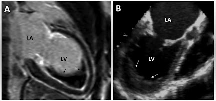
| Infarcts of most commonly affected organ |
Similar Posts -


No comments:
Post a Comment
Please do not enter any spam link in the comment box