Posts

Showing posts from December, 2018

Functions of skin

Functions of skin Introduction : skin is the largest organ of the body. It's thickness about 1-5mm according to location. Skin is made up following two layers... 1. Outer epidermis:   formed by the stratified epithelium. 2. Inner dermis: formed by the fibroblast, collagen fibers and histiocyte. Download book (physiology symbulingum ). Functions : Protective function: skin forms covering of all organs of the body and protects from Bacteria, Toxic substances, Mechanical blow and Ultraviolet rays. Sensory function: skin is considered as the largest sense organ of the body. It has many nerve endings which form specialized receptors. These receptors are stimulated by sensation of touch, pain, pressure and temperature convey these sensation to brain. Storage function: skin stores fat, water, chloride and sugar. Vitamin D3 : vitamin D3 is synthesized in skin by the action of ultraviolet rays on cholesterol. Regulation of body temperature:  Excess amount

Asphyxia

Aasgaduli.blogspot.com Asphyxia Definition : improper aeration of blood if continued for some time , then it produce a series of pathological changes and finally cause death which is known as Asphyxia.                                        OR Asphyxia is the condition characterized by combination of hypoxia and hypercapnea, due to obstruction of air passage. Codition when asphyxia occurs:  acute obstruction of air passage due to...    i. Strangulation .   ii. Hanging.  iii. Drowning. Stages of asphyxia: 1. Stage of  Hyperpnoea:   in this stage breathing becomes deep and rapid because of the powerful stimulation of respiratory centre by the excess carbon dioxide. Hyperpnoea is followed by dyspnea and cyanosis and eyes become more prominent. Duration of this stage extends only 1min. 2. Stage of convulsion or central excitation:  in this stage hypercapnea acts on brain and produce following effects...           i. Violent expiratory efforts.        

Hypoxia / Anoxia

Aasgaduli.blogspot.com Hypoxia / Anoxia Definition : Decrease oxygen level in blood or inadequate supply of oxygen to the tissues by the blood are known as Hypoxia. Causes :   Oxygen tension in arterial blood. Oxygen carrying capacity of blood. Velocity of blood. Utilization of oxygen by the cell. Classification : 1. Hypoxic hypoxia / Arterial hypoxia:   low oxygen tension in arterial blood. 2. Anemic hypoxia : low oxygen content of blood due to...                I. Lack of Hb.               II. CO poisoning.  3. Stagnant hypoxia / Hypokinetic hypoxia:   decrease rate of blood flow due to its velocity. 4. Histotoxic hypoxia : tissue are unable to use oxygen. Effects of hypoxia: 1. On nervous system -        i. Dullness.      ii. Dizziness.     iii. Confusion.     iv. Convulsion. 2. On digestive system:     i. Nausea.    ii. Vomitting.   iii. Vertigo.    iv. Loss of appitite. 3. On circulatory system:     i. Incr

Dyspnoea

Aasgaduli.blogspot.com Dyspnoea The term ''Dyspnoea" mean's difficulty in breathing. It is also known as Air hunger.             In normal condition breathing remains unnoticed, when this breathing is notice or comes in consciousness known as Dyspnoea. Definition : Difficulty in breathing called Dyspnoea. Causes:    Hypoxia Hypercapnia / Hypercarbia. Increase H+ ion concentration. Abnormality of diaphragm. Abnormality of chest wall. Failure of ventricles of the heart. Severe anaemia. Tissue lungs expiration. Pneumonia. Pulmonary edema. Poliomyelitis . Pneumothorax . Severe asthma. Download book ( physiology symbulingum)

Respiratory system (volumes & capacities)

Aasgaduli.blogspot.com Respiratory system  Volumes :  1. Tidal volume (TV) : TV is the volume of air breathed in and out of lungs in a single normal quiet respiration.  Normal value - 500ml. 2. Inspiratory reserve volume (IRV) : IRV is an additional volume of air that can be inspired forcefully after the end of normal inspiration . Normal value - 3300ml. 3. Expiratory reserve volume (ERV) : ERV is an additional volume of that can be expel out forcefully after normal expiration. Narmal value - 1000ml. 4. Residual volume (RV) : RV is a amount of air remain in the lungs after forceful expiration. Normal value - 1200ml. Capacities:  1. Inspiratory capacity (IC) : IC is the maximum volume of air that is inspired after normal expiration . It includes... IC (3800)= TV (500) + IRV (3300). Normal value - 3800ml. 2. Vital capacity (VC) : VC is the maximum volume of air that can be expel out forcefully after a deep inspiration . It includes.

Cell membrane

Cell membrane Definition : it is a protective sheath, and it is also known as plasma membrane, plasmalemma. This membrane separate ICF from ECF. it is a semipermeable membrane. Thickness of the cell membrane 75-111°A. Composition of cell membrane : Protein - 55% Lipid - 40% Carbohydrate - 5% Lipid layer of cell membrane :  Phospholipid. Cholesterol . Protein layer of cell membrane : 1. Integral protein - that pass through entire thickness of cell membrane from one side to other side. Example of integral protein:   Cell adhesion proteins. Cell junction protein. Some carrier or transport protein. Channel protein. Some hormones receptor. Antigens. Some enzymes. 2. Peripheral protein - are the partially embedded in the outer or inner surface of the cell membrane . Example of peripheral protein : Proteins of cytoskeleton . Some carrier or transport protein. Some enzymes. Functions of cell membrane : Protective function . Selecti

Cytoskeleton

Name :- Cytoskeleton Properties : It is an cellular organelle present throughout the cytoplasm. It is a complex network of structures with varying sizes. Cytoskeleton is consists of three major protein components i.e. microtubule, intermediate filaments and microfilaments. Microtubules : These are the straight, hollow and tubular structures.  These organelles are arranged in different bundles without the limiting any membrane.  Each tubules has 20-30 nano-meter diameter and length varies, it may be 1000 times more than the thickness.  Microtubules are formed by bundles of globular protein called  tubulin.  Tubulin has two subunits i.e. alpha and beta. Intermediate Filaments (IF) : IF form a network around the nucleus and extend to the periphery of the cell. Diameter of each filament is about 10 nano-meter. IF are formed by rope-like protein polymers (fibrous proteins) IF are divided into five subclasses :- Keratins (in epithelial cells) Glial flaments (in astrocytes) Neurofilaments (in

Ribosomes

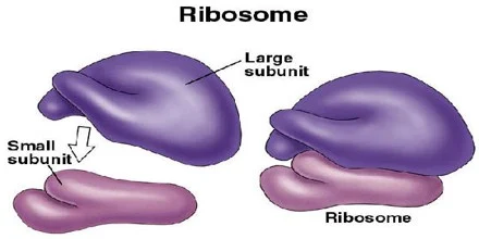
Name : - Ribosomes , protein factories Properties These are the dot like or granular structure without limiting any membrane. It's diameter 15nm. It is made up of 35% protein and 65% ribonucleic acid (RNA) called ribosomal RNA. Human ribosomes are 80S ('S' for Svedberg's Unit) Each ribosome has two subunits i.e. larger (60S) and smaller (40S) Types of Ribosomes : Ribosomes attach to the endoplsamic reticulum called rough endoplasmic reticulum (RER). Free ribosome - that are distributed in the cytoplasm. Function of Ribosomes :-   Rough endoplasmic reticulum :- Synthesis of protein such as enzymatic protein, hormonal protein, lysosomal protein and the protein of the cell membrane. Free ribosomes :- are responsible for synthesis of protein in haemoglobin, peroxisome and mitochondria. Ribosomes Download book ( physiology symbulingum) Similar Post :-  Mitochondria Lysosomes Rough Endoplasmic Reticulum Cytoskeleton Cell membrane Golgi apparatus

Endoplasmic reticulum

Name :- Endoplasmic Reticulum (ER) Properties : ER is a network of tubular and microsomal vesicular structures which is present inside the cell. The tubules and vesicles are interconnected. ER covered by a limiting membrane which is formed by proteins and bilayered lipids. The lumen (diameter - 400 to 700 angstrom) of ER contains a fluid medium called endoplasmic matrix . Total surface area of ER (in some cells like liver cells) can be as much as 30-40 times the cell membrane area. ER forms the link between nucleus and cell membrane by connecting them. Types of ER .... Rough Endoplasmic Reticulum (RER) / Granular Endoplasmic Reticulum (GER) Smooth Endoplasmic Reticulum (SER) / Agranular Endoplasmic Reticulum (AER) / Tubular Endoplasmic Reticulum (TER) 1. RER : Rough appearance of ER is due the attachment of ribosomes (granules) to it's outer surface. Hence, it is also celled GER. Functions :  Synthesis of proteins (specially those proteins which are secreted from the cells like ins

Lysosomes

Name :- Lysosomes, garbage system of the cell, suicidal bag of the cell Properties : These are membrane bound vesicular structures founds throughout the cytoplasm. These are formed by the process of packaging in the golgi apparatus. Enzymes of lysosomes are synthesized in rough endoplasmic reticulum. Among the organelles, the lysosomes have the thickest covering membrane that is formed by a bilayered lipid material . Lysosomes have 50 different hydrolytic enzymes, known as acid hydroxylases. Types of Lysosomes : Primary lysosomes, this is an inactive of form of lysosomes. Secondary lysosomes, this is an active form of lysosomes (lysosomes active during phagosome or endosome and at this time lysosomes pH become acidic and enzymes are activate). Lysosomal Enzymes :   Proteases :- which hydrolyze the proteins into amino acids. Lipases :- which hydrolyze the lipids into fatty acids and glycerides. Amylases :- which hydrolyze the polysaccharides into glucose. Nucleases :- whi

Mitochondria

Name :  Mitochondrion ( plural - mitochondria), power house of the cell. Properties :  It is membrane bounded cytoplasmic organelle. It's shape rod or oval, and diameter 0.5 - 1.0 micrometer. It covered bilayered membrane  Outer membrane : is smooth and encloses the contents of mitochondrion as well various enzymes such as acetyl-CoA synthetase, glycerophosphate, acetyltranferase. Inner membrane : is folded in the form of shelf-like inward projection called cristae (crista). Cristae contain many enzymes and other protein molecules which helps in functioning. (Functions + enzymes + protein molecules in cristae = respiratory chain or electron transport chain). Enzymes of respiratory chains are 4-8 in list below.   It's DNA contain 37 gene. It responsible for maternal child relation. It contains many enzymes and protein. Increase no of mitochondria by the exercise. It is the site of aerobic respiration Download book ( physiology symbulingum) Enzymes and protein :

Hemophilia

Aasgaduli.blogspot.com Hemophilia  Definition - it is a congenital disease and disorder of secondary haemolytic disease. Cause :- congenital defect of factor VIII (anti hemophlic factor). Clinical features :-  Disease is uncommon in indians. After injury bleeding stops. Bleeding reappears after few minutes. Continuous prolonged bleeding. Treatment :- Transfusion of blood. Transfusion of plasma. Transfusion of anti hemophilic factor. Download book ( physiology symbulingum)

Mast cells

Aasgaduli.blogspot.com Mast cells  Mast cell present in lungs . It is a large cell and looks like basophills. They are hypersensitive to allergy and in some other conditions. Mast cell secrets :- 1. Heparin (anti coagulant). 2. Histamine ( anti allergic). 3. Serotonin. 4. Hydrolytic enzyme. Download book ( physiology symbulingum)

Clotting factors

Aasgaduli.blogspot.com Clotting or coagulation factor : Fibrinogen. (I) Prothrombin. (II) Thromboplastin or tissue factor. (III) Calcium. (IV) Labile factor. (V) Absent... (VI) Stable factor. (VII) Antiheamophlic factor. (VIII) Christmas factor (IX) Stuart factor. (X) Plasma thromboplastin anticedent. (XI) Hageman factor. (XII) Fibrin stablizing factor. (XIII) Download book ( physiology symbulingum)

Functions of Platelets

Aasgaduli.blogspot.com Functions of Platelets : 1. Help in blood clotting. 2. Haemostasis of blood. 3. Prevent clot retraction. 4. Release various substances. 5. Repairing of capillary endothelium. Download book ( physiology symbulingum)

Functions of W.B.Cs

Aasgaduli.blogspot.com Functions of W.B.C. Functions of W.B.Cs 1. Phagocytosis : when bacteria and forign particles invade body, then leukocyte comes out from blood and their psedopodia process engulf and destroy them with the help of hydrolytic enzyme. 2. Antibody formation : lymphocyte are manufacture Beta and Gamma globulin. Which is a part of antibody. 3. Formation of fibroblast: leukocytes converts into fobroblast at the area of inflammation and helps in repair. 4. Manufacture of trephones : they prepare some substance from plasma proteins which have great influence on nutrition, growth and repair. These substance are known as trephones. 5. Secretion of heparin : Basophil secrets heparin which prevents from intravascular clotting. 6. Antihistamine  function : eosinophil have antihistamine or anti allergic function. Download book ( physiology symbulingum)

Gastric juice

Gastric juice 1. Types of Gastric gland 2. Properties. 3. Composition. 4. Secretion. 5. Factor responsible. 6. Functions. 1. Types of gastric glands :  Glands depends upon the situation - A. Fundic/ main/proper/oxyntic gland : it secrets 1. HCL 2. Mucin 3. Pepsinogen 4. Intrinsic factor B. Cardic gland : it secrets 1. Mucin 2. Pepsinogen C. Pylorus gland : it secrets a solution which is rich in mucus and don't contain HCL. It also secret gastrin. Types of cell of gastric glands :  1. Chief or peptic cell - it produce Renin, pepsin. 2. Oxyntic or parietal cells - it produce HCL. 3. Neck mucus cells - It produce Mucus, Pepsinogen. 4. Surface epithelial cells - it produce Mucus. 5. Stem cells - it produce G- cell or Daughter cell. 6. G- cells - it produce Gastrin Naerve supply - Vagus nerve 2. Properties :  Volume                   - 1200 - 1500ml/day Reaction                 - highly acidi

Transport across the cell membrane

Transport across the cell membrane The fluid which is present inside the cell membrane  known as intracellular fluid (ICF), and the fluid which is present out side the cell is known as extracellular fluid (ECF).                   The cell membrane is present between ICF and ECF. This cell membrane is semipermeable and it allows selective molecules through it. Transport across cell membrane  Process :  the movement of substances across cell membrane by following two process... 1. Passive transport 2. Active transport Passive transport : use own energy , no ATP use. Types :  1. Diffusion  : Definition - a process in which solute move from higher concentration area to lower concentration area known as diffusion. Diffusion are two types... A. Simple diffusion : in which a. Lipid layer of cell membrane which is known as simple lipid diffusion. This layer is permeable to lipid soluble substances like oxygen and carbon dioxide. b. Protein layer of cell mem

Functions of gall bladder

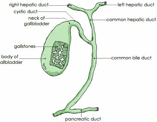
Functions of gall bladder a. The bile secreted from liver and stored in gall bladder.  b. Gall bladder is not essential for life.  c. The removal of gall bladder called cholecystectomy.  d. After cholecystectomy ,patients does not suffer from any major disadvantage.  e. In some species gall bladder is absent. Functions: - 1. Storage of bile. 2. Alteration of pH of bile. 3. Secretion of mucin. 4. Maintenance of pressure in biliary system. Gallbladder  Download book ( physiology symbulingum)

Cell

Cell Cell means small room or a chamber. Cell word was given by Robert Hook in 1663 and cell discovered also in 1663 by Robert Hook. Definition : cell are the structural and functional unit of a living body. These are the smallest unit which contains all the features of life. General features of cell :   1. Cell required Oxygen, nutrition for their survival. 2. Cell can produce own energy itself, which is important for their Growth, Repair, Activities. 3. Cell produce carbon dioxide and other metabolic waste products during his activities. 4. Cell shows immediate response against bacteria, viruses or toxic substances entering in the body. 5. Cell reproduced through cell division except neurons and cardiac muscle fibers. Structure of a cell : a cell contains... Body Cell membrane 1. BODY  Nucleus Cytoplasm     A. Nucleus : it is covered by a double layer membrane known as nuclear membrane. Nucleoplasm : it is a gel like substance

Anterior abdominal wall

Anterior abdominal wall Anterior abdominal wall is made up by the skin, ( outer to inner) superficial fascia, muscles, nerves, lymph, vessels. It starts from 9 T.V. to pubic symphysis. Blood supply :  Epigastric artery, Deudenal artery and superficial circumflex. Download  book:  Anatomy vol-2 BD chourasia (Lower limb, Abdomen and pelvis ). Muscles of anterior abdominal wall - 1. External oblique abdominis - Origin : external surface & inferior border of the ribs with eight slips. Insertion : xiphoid process, linea Alba, pubic symphysis, pubic crest, pectineal line of the pubis, iliac crest. Action : forward bending, rotation, abduction. Nerve supply : T 7 - T 12 2. Internal oblique muscle - Origin : inguinal ligament 2/3, iliac crest 2/3, thoracolumbar fascia. Insertion : 10-12 ribs Action : bilateral contraction, compress the abdomin, unilateral contraction, ipsilaterally rotates the torso. Nerve supply : T 7 - T 12 , bran

Abdominal regions

Abdominal region The abdomen is a large area so for the study purpose it split into nine region by two horizontal plane and two vertical plane. Horizontal plane -  1. Transpyloric plane : a horizontal line, halfway between xiphoid process and the umblicus, passing through the pylorus of the stomach. 2. Inter tubercular plane : a horizontal line which joined to iliac crest each other. Vertical plane - This plane run form the mid clavicle to the mid inguinal point these plane are mid clavicular line. That is two in no right and left. Download  book: Anatomy  vol-2 BD chourasia ( Lower limb, Abdomen and pelvis ).  Regions - 1. Right hypogastric region - Viscera : Liver, gallbladder.     Abdominal regions 2. Epigastric region - Viscera : stomach, duodenum, pancreas, liver. 3. Left hypochondric region - Viscera : stomach, spleen, left colic flexure, Liver. 4. Right lumbar region - Viscera : Right kidney, ureter, ascending colon. 5. Umblica