Posts

Showing posts from February, 2019

Femur

Image
Femur The femur is a latin word it means thigh. Femur is the longest and strongest bone of the body. Femur has 27% length of total body length. Weight of femur is about 380gm in males and 279gm in females. Quantity of femur are two in number. Side Determination 1. Head directed medially. 2. Medial and lateral condyle (lower end) inferiorly. 3. Cylindrical shaft is convex forward. Joint Formation  1. Hip joint - between head of femur and acetabulum of Hip bone . 2. Knee joint - between lower end of femur and upper end of Tibia. Features  It has upper end, lower end and shaft. Right femur anterior aspect Upper end It has head, neck, greater trochanter ( Greek- runner), lesser trochanter, intertrochantric line and intertrochantric crest. Head -  It forms more than half a sphere and is directed madially, upwards and slightly forwards. It articulates with the acetabulum to form hip joint. It has a pit that is called fovea.
Digestion of lipid

Image
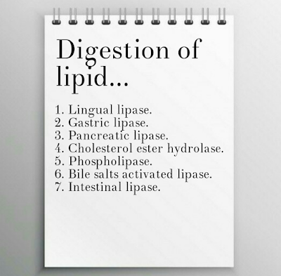
Digestion of lipid Definition of digestion Brocken down of big food particles into small food particles, which can be absorbed and use as a nutrients for the body. Classification of lipid 1. Simp;e fat - a. Triglyceride / Neutral fat  b.  cholesterol . 2. Compound fat - a. Phospholipid b. Glycolipid c. Lipoprotein 3. Derived fat - a. Prostaglandin b. Leuketrin We are lipid intake with food in the form of triglyceride mostly while some parts of cholesterol and cholesterol ester. The enzyme which help in digestion of lipid called lipolytic  enzyme.    Digestion of lipid in mouth Saliva contain a lipolytic enzyme called lingual lipase, but this enzyme not act on food in mouth and engulfed with food. Digestion of lipid in stomach Gastric juice contain a lipolytic enzyme called gastric lipase, which act on triglyceride and converts into fatty acids and glycerol. Digestion of lipid in small intestine Intestine have two ju

General anatomy (Handbook) by BD Chourasia

Image
General anatomy (Handbook) by BD Chourasia General anatomy (Handbook) by BD Chourasia Download book pdf About file: - File type - pdf File size - 20.76 MB Pages - 276 Language - English

Digestion of protein

Image
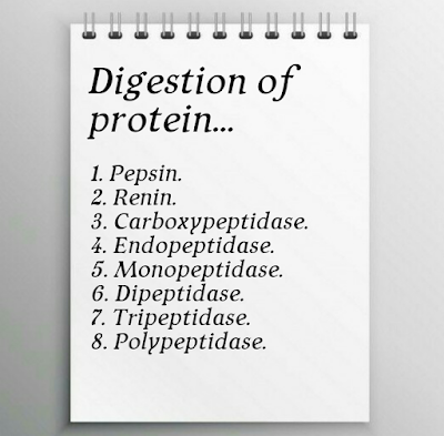
Digestion of protein Definition Broken down of big food particles into small food particles by the chemical process, which can be absorbed and use as a nutrients. The enzyme which help in protein digestion called proteolytic enzyme. Enzymes of protein digestion Digestion of protein in mouth Saliva secrets in the mouth which don't have any proteolytic enzymes, so protein digestion not occur in the mouth. Protein digestion in stomach Gastric juice contain a proteolytic enzyme called pepsin. Pepsin act on protein and converts into protease, peptone and polypeptide. pepsin act in highly acidic medium. Digestion of protein in small intestine Small intestine contain two juices... 1. Pancreatic juice 2. Succus entericus Pancreatic juice Pancreatic juice have carboxy peptidase and endo-peptidase ( trypsin and chymotrypsin ) enzymes, these enzyme are proteolytic enzyme. Trypsin act on protein and convert into di-peptide. Chymotrypsin act on pepton

Digestion of carbohydrate

Image
Digestion of carbohydrate Definition Brockendown of  big food particles into small food particles by the chemical process, which can be absorb and use as a nutrients. The enzyme which help in digestion of carbohydrate called amylolytic enzyme. Classification of carbohydrates 1. Monosaccharide - Hexoxes ( glucose ) , Pantoses. 2. Disaccharide - Sucrose ( Glucose + Fructose ), Lactose ( Glucose + Galactose ) 3. Polysaccharide - Starch, Glycogen, Amylose. Enzymes of digestion of carbohydrate Digestion in mouth Mouth secrets saliva which contain salivary amylase/ ptylin /ptyalin . This enzyme act as amylolytic enzyme. This enzyme acts on starch and converts into dextrose and maltose. Ptyalin inactive in highly acidic medium, so when food reach in stomach then ptyalin is inactive due to increase acidity. Digestion in stomach   Gastric juice contain a enzyme called gastric amylase , which is a very weak enzyme so it's function is negligible in stomach.

Cardiac cycle

Image
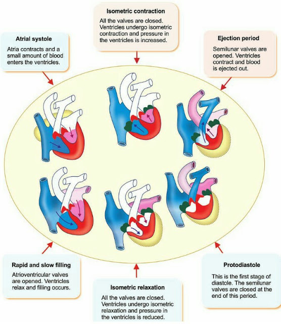
Cardiac cycle Definition The change occur in heart during one heart beat, this process continue repeated in sequence manor called cardiac cycle. Duration of cycle Heart rate - 70 - 80 / min. Average - 75 /  min. One heart beat - 60/75 = 0.8 sec. Each cardiac cycle is starts from spontaneous generation of impulses by SA node, this impulse travel from SA node to AV node through intermodal fibers than bundle of his than right and left bundle branch than purkinge fibers than it distributes all myocardium. Events of cardiac cycle Events of cardiac cycle Atrial events ( 0.8 sec ) 1. Atrial systole  ( 0.1 sec ). 2. Atrial diastole ( 0.7 sec ). Ventricular events ( 0.8 sec ) 1. Ventricular systole ( 0.3 sec ). 2. Ventricular diastole ( 0.5 sec ). Ventricular systole ( 0.3 sec ) 1. Isovolumetric phase ( 0.05 sec ). 2. Rapid ejection phase ( 0.1 sec ). 3. Reduce ejection phase ( 0.15 sec ). 1. Iso-volumetric phase ( 0.05 sec ) Valve - close al

Mediastinum

Image
Mediastinum It is the medial septum of the thorax between two lungs and include mediastinal pleura. Boundaries of mediastinum Anteriorly - Sternum. Posteriorly - Vertebral column. Superiorly -Thoracic inlet. Inferiorly - Diaphragm. On each side - Mediastinal pleura. Subdivisions of the mediastinum Division of mediastinum Mediastinum divided into two parts... 1. Superior mediastinum 2. Inferior mediastinum inferior mediastinum further divided into 3 parts... a. Anterior mediastinum b. Middle mediastinum c. Posterior mediastinum Superior Mediastinum Boundaries Anteriorly - Manubrium sterni. Posteriorly - Upper four thoracic vertebra. Superiorly - Thoracic inlet. Inferiorly - An imaginary plane ( passing through the sternal angle and the lower border of 4th thoracic vertebra). On each side - Mediastinal pleura. Contents 1. Trachea and oesophagus. 2. Muscles - Origin of ...(i. Sternohyoid, ii.Sternothyroid, iii. Lower end of longus colli

Clavicle

Image
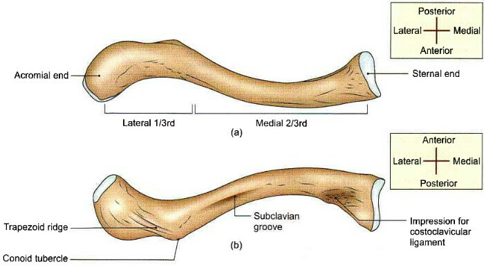
Clavicle  Synonames Collar bone, Beauty bone. Anatomical position It extends laterally and horizontally at the root of the neck Side Determination  1. Flattend end laterally. 2. Medial two-third of clavicle is convex anteriorly. 3. Rough area on the lateral side faces inferiorly. Special Features  It is only long bone which lies horizontally. It has no medullary cavity (Bone marrow). It is the first bone to ossifies in the body ( second bone to ossifies Mandible). Almost whole clavicle visible through skin can be palpable. It has the only long bone which has two primary center of ossification. It has only long bone which ossifies in membrane. General features of right clavicle (a) Superior aspect (b) Inferior aspect Features It has 2 ends (Medial or Sternal, Lateral or Acromial end) and a shaft. Lateral end  It is flattend from above downwards. It articulate with the acromian process of scapula to form acromioclavicular join

Scapula

Image
Scapula Type -  Flat Quantity - 2 in numbers Location  It is posterolateral aspect of the thorax, against 2-7th rib. Side Determination Glenoid cavity looks laterally. Coracoid process is directed forwards anteriorly. Spinous process always posteriorly. Features Surfaces  1. Costal Surface 2. Dorsal Surface Costal Surface It is also called as subscapular fossa. It is concave. Costal surface of right scapula Dorsal Surface A spine divide it (Dorsal Surface) into two parts... 1. Supra Spinous fossa or Surface. 2. Infra Spinous fossa or Surface. Dorsal surface of right scapula Borders 1. Superior Border. 2. Lateral Border. 3. Medial Border Superior Border It is thin and shortest  border. Suprascapular notch present on it. Lateral Border It is the thickest border. Medial Border It is also called as vertebral border. Angles 1. Superior Angle. 2. Inferior Angle. 3. Lateral