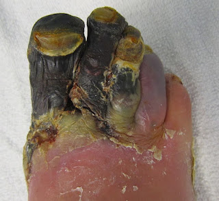
Posts

Showing posts from March, 2020

Necrosis

Image
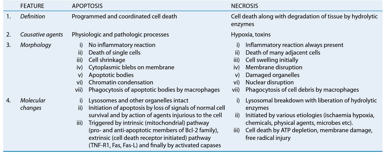
Necrosis NECROSIS – Pathological cell or cells death in living body due to injury. NECROBIOSIS – It is a physiological cell death e.g. desquamation of surface epithelium. APOPTOSIS – It is a programmed cell death, it may be pathological or physiological. In this process the nucleus is condensed the cell is divided into small membrane bounded bodies called apoptotic bodies. Eventually the apoptotic bodies are engulfed by phagocytes. Causes – 1. Hypoxia (lack of O 2 or ischaemia) – disturbed blood supply. 2. Physical agents – Excessive heat and cold etc. 3. Chemical agents – Strong acids and alkaloids etc. 4. Immunological reactions General features – 1. The cytoplasm is homogeneous or granular and eosinophilic. 2. The cell cytoplasm may show vacuolation. 3. The nucleus shows three types of changes …             a. Pyknosis – It is a initial change, in which nuclear material is condensed.             b. Karyorrhexis – The nucleus is divided into small fra

Gangrene

Image
Gangrene Massive cell death of a part of living tissue superadded putrefaction due to saprophytic bacteria. Classification  1. Dry gangrene 2. Wet or moist gangrene 3. Gas gangrene 1. Dry Gangrene : This type of gangrene is seen in that part of the body there is lack of water quantity like - hands and feet. Causes :   Gradual vascular obstruction as seen in senile gangrene, diabetic gangrene. Sudden vascular obstruction as seen in thrombosis, embolism and ligation. Extreme cold (frost bite). Strong acid, coagulate the fluid of the tissue so produce dry gangrene while strong alkalies liquefies the tissue so produce the moist gangrene. Features of dry gangrene :   This type of gangrene is seen in the body there is lack of water like hands and feet. This type of gangrene generally starts in distal part (great toe) and spread toward the heart. Growth rate of this type gangrene is slow. The affected part is initially waxy then becomes dry and shrunke

Atrophy

Image
ATROPHY Definition – Reduction of the number and size of parenchymal cells of a normal organ or its parts called atrophy. HYPOPLASIA – Incomplete developments of a tissue or organ. APLASIA – Defective development or congenital absence of a tissue or organ. Causes – It may be physiological or pathological… PHYSIOLOGICAL ATROPHY – Atrophy of lymphoid tissue with age. Atrophy of thymus in adult life. Atrophy of gonads after menopause. Atrophy of brain with ageing. Osteoporosis with reduction in size of bony trabeculae due to ageing. PATHOLOGICAL ATROPHY – Starvation atrophy – This type of atrophy is due to lack of food and using stored food. It is seen in cancer and severely ill patients. Ischaemic atrophy – Gradual diminution of blood supply due to atherosclerosis may result in shrinkage of the affected organ e.g. small atrophic kidney and atrophy of the brain due to atherosclerosis. Disuse atrophy – Prolonged diminished functional activity may cause atroph

Cell injury

Image
Cell injury Definition : Whenever cell adaptation exceed due to disturbance of cell function called cell injury. Causes of cell injury :  Hypoxia and ischaemia (common cause) - Deficiency of oxygen or lake blood supply or ischemia.  Physical agents - Include excessive heat, excessive cold, trauma, electricity, radiations, sudden change in atmospheric pressure. These are the physical agents that caused cell injury. Chemical agents - Include strong acid, strong alkalides, drugs, pollutants. These are the chemical agents that caused cell injury. Microbial agents - Includes virus, bacteria, fungi and some protozoa. These are the microbial agents that caused cell injury. Immunological reactions - Hypersensitivity reactions, anaphylactic reactions and autoimmune diseases. Genetical effects Aging  Nutritional imbalance - Lake ( starvation, marasmus, kwashiorkor, anaemia) or exceed (obesity, atherosclerosis, heart disease and hypertension) of nutrition.  PATHO

Hypertrophy Of The Heart

Image
HYPERTROPHY OF THE HEART Definition -   Hypertrophy of the heart is defined as an increase in size and weight of the myocardium. In which involve predominantly the left or the right or both sides of the heart. It generally results from increase pressure load while increased volume load results in hypertrophy with dilatation. It is a compensatory mechanism of the heart. Common causes – (usually causes are unknown) Stretching of myocardial fibrosis. Anoxia (e.g. in coronary atherosclerosis). Certain hormones (e.g. Catecholamines and pituitary growth hormone). Left ventricular hypertrophy. Causes of left ventricular hypertrophy – Systemic hypertension. Aortic stenosis and insufficiency / regurgitation. Mitral insufficiency / regurgitation. Coarctation of the aorta. Occlusive coronary artery disease. Congenital anomalies like septal defects and patent ductus arteriosus. Conditions with increased cardiac output e.g. thyrotoxicosis, anaemia, arteriovenous fi

COVID-19

Image
COVID-19 (Coronavirus Disease 2019) Definition - It is an acute highly infectious viral disease caused by SARS cov-2. It affects only respiratory system. Cases may be asymptomatic to severe and critical, 80% cases recover spontaneously and 15-20% cases are severe and critical and characterized by fever, non-productive cough, myalgia shortness of breath and  may show severe pneumonia even death. Agent factors - AGENT - novel coronavirus (SARS cov-2) RESISTANCE - It resists at surface from few hours to several days depending upon the condition (e.g. temperature and humidity of the environment) Novel coronavirus SOURCE OF INFECTION - Case - clinical and subclinical (80 % of infections are mild or asymptomatic, 15 % are severe infection and 5 % are critical infection) (as per on that days data) INFECTIVE MATERIAL - Oropharyngeal secretion, nasal secretion (research going on) PERIOD OF COMMUNICABILITY - Starts before 24-48 hours appearance of symptoms till patient r

Diphtheria

Diphtheria Definition -  It is an acute infectious bacterial disease caused by corynebacterium diphtheriae found in clinical forms i.e. Anterior nasal, Pharyngotonsiller (faucial) and Laryngotracheal. The bacteria produces a powerful exotoxin locally and it is responsible for the formation of pseudo-membrane, local oedema and lymphadenopathy with sign of toxaemia. Agent factors - AGENT -  Corynebacterium diphtheria. It is a gram +ve non-motile organism and it has no invasive power but produce a powerful exotoxin. There are two types of strains –  (a) Pathogenic strain – Gravis, mitis, belfanti and intermedius.  (b) Non-pathogenic strain – They are capable to change into pathogenic strains due to ß-phase. INFECTIVE MATERIAL – Nasopharyngeal secretion and contaminated wounds, fomite and dust. SOURCE OF INFECTION -  (a) Case (5%) – mild and frank clinical cases. (b) Carrier (95%) – Temporary and chronic carrier PERIOD OF COMMUNICABILITY – 14-28 days from onse