Posts

Showing posts from December, 2019

Williams GYNECOLOGY

Image
Williams GYNECOLOGY CONTENTS Editors Contributors Artists Preface Acknowledgments SECTION I : BENIGN GENERAL GYNECOLOGY 1. Well Woman Care 2. Techniques Use For Imaging In Gynecology 3. Gynecologic Infections 4. Benign Disorders Of The Lower Genital Tract 5. Contraception And Sterilization 6. First Trimester Abortion 7. Ectopic Pregnancy 8. Abnormal Uterine Bleeding 9. Pelvic Mass 10. Endometriosis 11. Pelvic Pain 12. Breast Disease 13. Psychological Issue And Female Sexuality 14. Pediatrics Gynecology SECTION II : REPRODUCTIVE ENDOCRINOLOGY, INFERTILITY AND THE MENOPAUSE 15. Reproductive Endocrinology 16. Amenorrhea 17. Polycystic Ovarian Syndrome and Hyperandrogenism 18. Anatomic Disorders 19. Evaluation Of the Infertile Couple 20. Treatment Of the infertile Couple 21. Menopausal Transition 22. The Mature Woman SECTION III : FEMALE PELVIC MEDICINE AND RECONSTRUCTIVE SURGERY 23. Urinary Incon
Review of PHARMACOLOGY

Image
Review Of PHARMACOLOGY CONTENTS History Of Pharmacology General Pharmacology Autonomic Nervous System Autacoids Cardiovascular System Kidney Endocrinology Central Nervous System Anaesthesia Hematology Respiratory System Gastrointestinal Tract Chemotherapy A : General Consideration and Non-specific Antimicrobial Agents Chemotherapy B : Antimicrobial for specific Conditions Chemotherapy C : Antineoplastic Drugs Immunomodulators Other Topics and Advers Effects Drugs of Choice New Drugs Recent Topics Latest Papers Image Based Qustions Review of PHARMACOLOGY ABOUT BOOK Name Book Review of PHARMACOLOGY Author Gobind Rai Garg & Sparsh Gupta Type Pdf Size 22.7 MB Pages 785 Pages type Colored Quality Good Related Posts Textbook of Pharmacology and Therapeutics Pharmacology Books

Appendicitis

Image
Appendicitis Acute inflammation of the appendix. It is seen most commonly in older children and young adults, and it is uncommon in extremes of age. Etiopathogenesis : Most often cause of appendicitis is low bulk or cellulose and high protein diet intake. Other cause of appendicitis are obstructive and non-obstructive - A. Obstructive :  Faecolith Calculi Foreign body Tumour Worms (especially Enterobius vermicularis) Diffuse lymphoid hyperplasia, especially in children. B. Non-obstructive : Haematogenous spread of generalised infection Vascular occlusion Inappropriate diet lacking roughage Appendicitis You may also read - Dysentery MORPHOLOGICAL FEATURES Grossly :  1. Acute appendicitis (early) -          i. The organ is swollen          ii. Serosa shows hyperaemia. 2. Acute suppurative appendicitis (well-developed) -          i. The serosa is coated with fibrinopurulent exudate.         ii. Engorged vessels on the surface. 3. Ac

Dysentery

Image
Dysenteries Definition : Diarrhoea with abdominal cramps, tenesmus and mucus in the stool called dysentery. CLASSIFICATION  Bacillary dysentery Amoebic dysentery 1. BACILLARY DYSENTERY : It is the infection of Shigella species (S. dysenteriae, S. flexnery, S. boydii and S. sonnei). Etiopathogenesis : Infection of the shigella species occurs by faeco-oral route with contaminated food and water. Infection is seen commonly in poor personal hygiene persons and overcrowding areas. The housefly play a major role in spread the infection. MORPHOLOGICAL FEATURES Grossly :  The lesions are mainly found in the colon sometime in the ilium. Superficial transverse ulcerations of mucosa of the bowel wall occurs in the region of lymphoid follicles and perforation maybe seen. The intervening mucosa is hyperaemic and oedematous. All of these recover completely after acute attack. Microscopically :  Mucosa of the lymphoid follicles are necrosed. The sur

Typhoid Fever

Image
Typhoid / Enteric Fever Enteric fever is an acute infection of the salmonella typhi (typhoid fever) or salmonella paratyphi ( paratyphoid fever) and may be food poisoning . ETIOPATHOGENESIS : The typhoid bacilli are ingested through contaminated food and water. Initially it is asymptomatic and incubation period is about 2 weeks. The bacilli invade the lymphoid follicles and peyer's patches of small intestine and proliferate. MORPHOLOGICAL FEATURES : The lesions are observed in intestine and many other organs - 1. Intestinal lesions Grossly : Terminal ilium is affected mostly but lesions may be seen in the jejunum and colon. Peyer's patches show oval typhoid ulcers their long axis along the length of the bowel. The base of the ulcers is black due to sloughed mucosa. The margins of the ulcers are slightly raised due to oedema and cellular proliferation. The regional lymph nodes are enlarged. Microscopically : There is hyperaemia, oedema and cellular prolifera

Surgery ( Exam Preparatory Manual for Undergraduates)

Image
Surgery ( Exam Preparatory Manual for Undergraduates) CONTENTS SECTION 1 GENERAL SURGERY Wound Healing Fluid Electrolyte and Acid-Base Imbalance Metabolism and Nutrition Blood Transfusion and DIC SIRS, Shock and MODS Basic of Surgical Technologies and Advanced Surgery Surgical Infections Trauma and Damage Control Surgery Perioperative Surgery Surgery of the Salivary Glands Miscellaneous General Surgery Topics of Importance Medicine in Surgery SECTION 2 GASTROINTESTINAL, HEPATOBILIARY AND PANCREATIC SURGERY Esophagus and Diaphragm Stomach and Duodenum Small Intestine Large Intestine Appendix Rectum and Anal Canal Liver Gallbladder and Bile Duct Pancreas Spleen Hernias Gastrointestinal Oncosurgery Miscellaneous SECTION 3 GENITOUROLOGY  Kidney and Ureter Urinary Bladder Prostate Testis and Scrotum Penis and Urethra Uro-oncology SECTION 4 NEUROSURGERY SECTION 5 CARDIOTHORACIC AND VASCULAR SURGERY Cardiac and Thoracic Surgery

Influenza

Influenza / Bird flue / Swine flue Definition : It is an acute highly infectious viral disease caused by influenza virus A, B and C. Characterised by high grade fever associated with malaise, headache, chills, sneezing, coryza etc. AGENT FACTORS Agent : Influenza virus A, B & C ( arthomyxoviridae family), antigenically different, no cross immunity. Type A & B viruses has antigen (Haemagulutinin and Neuraminidase) Haemagulutinin (H antigen) - have 16 subtypes H 1 , H 2 , H 3 ….H 16 .  It provide attachment of the virus to susceptible host. Neuraiminidase ( N antigen) - have 9 subtypes N 1 , N 2 , N 3 ….N 9 .  It is responsible for the release of the virus from the infected cells Source Of Infection : Case - Clinical and Subclinical. Infective Materials : Nasopharyngeal secretion. Period Of Communicability : 1-2 days before & 1-2 days after the appearance of symptoms. HOST FACTORS Age : All ages are susceptible. S

SABISTON TEXTBOOK of SURGERY

Image
SABISTON TEXTBOOK of SURGERY The BIOLOGICAL BASIS of MODERN SURGICAL PRACTICE About Book Name book SABISTON TEXTBOOK of SURGERY (20th edition Author Beauchamp, Evers and Mattox Type pdf Size 366.6 MB Pages 2136 Page Type Colored Quality Good Sabiston Textbook of Surgery Download Book From Here

Textbook of Pharmacology & Therapeutics

Image
Textbook of Pharmacology & Therapeutics CONTENTS FOREWORD PREFACE ACKNOWLEDGEMENT PART I GENERAL PRINCIPLES Introduction to therapeutics Mechanism of drug action (Pharmacodynamics) Pharmacokinetics Drug absorption and routes of administration Drug metabolism Renal excretion of drugs Effects of disease on drug disposition Therapeutic drug monitoring Drugs in pregnancy Drugs in infants and children Drugs in the elderly Adverse drug reaction Drug interaction Pharmacogenetics Introduction of new drugs and clinical trials Cell based and recombinant DNA therapies Alternative medicines : herbals and nutraceuticals PART II THE NERVOUS SYSTEM Hipnotics Schizophrenia and behavioural emergencies Mood disorders Mood disorders and degenerative CNS disease Anti epileptics Migraine Anaesthetics and muscles relaxants Analgesics and the control of pain PART III THE MUSCULOSKELETAL SYSTEM Anti inflammatory drugs and the treatment of arthritis

Acute Peptic Ulcers

Peptic Ulcers Definition : Peptic ulcers are the areas of degeneration and necrosis of gastrointestinal mucosa exposed to acid peptic secretions. They occur most commonly (98-99%)  in either the duodenum or the stomach in the ratio 4:1. It may be acute or chronic. Acute Peptic Ulcers / Acute Stress Ulcers  Definition : Acute peptic ulcers are multiple, small mucosal erosions, seen most commonly in the stomach but sometime involve duodenum also. Etiology : Most common cause of ulcers are stress - 1. Psychological stress 2. Physiological stress -  Shock Severe trauma Septicemia or sepsis Extensive burns (Curling's ulcers in the posterior aspect of the first part of the duodenum) Intracranial lesions ( Cushing's ulcers developing from hyper-acidity due to excessive vegal stimulation)  Drug intake (aspirin, steroids, butazolidine, indomethacin) Local irritants (alcohol, smoking, coffee ) Pathogenesis : Mucosal digestion from hyper-acidity (impo

Mumps (Parotitis)

Mumps / Parotitis Definition : It is an acute highly infectious viral disease caused by a myxovirus characterised by one or both parotid gland swelling and sometime associated with constitutional symptoms like fever, headache, malaise etc. AGENT FACTORS Agent : Myxovirus Parotiditis ( it is a RNA virus of the myxovirus family). These virus has affinity with glandular and nervous tissue. These virus can affects parotid gland. testes, ovaries, pancreas and nervous system. Source Of Infection : Case - clinical and subclinical ( 30-40% of all cases). Infective Materials : Oropharyngeal secretions, Saliva, Blood, Milk and CSF. Period Of Communicability : 4-5 days before onset of symptoms and till swelling subside. HOST FACTORS Age : It can affects in any age but usually age is 5-15 years (severe in adults). Sex : Equal effects on both sexes. Immunity : Life long immunity, after single attack. Secondary Attack Rate : 86% ENVIRONMENTAL FACTORS Season : Throughout the

Review of PATHOLOGY and Genetics

Image
Review of PATHOLOGY and Genetics CONTENTS  Cell Injury Inflammation Hemodynamics Genetics Neoplasia Immunity Anemia and Red Blood Cells White Blood Cells and Platelets Cardiovascular System Respiratory System Kidney and Urinary Bladder Gastrointestinal Tract Liver Genital System and Breast Central Nervous System Endocrine System Musculoskeletal System Miscellaneous Recent Papers Important Stains and Bodies Image Based Questions

Emphysema

Image
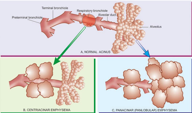
Emphysema Definition It is the permanent dilatation of air spaces and destruction of their wall distal to terminal bronchiole. Classification of emphysema 1. Centriacinar/ centrilobular/ proximal part emphysema . 2. Panacinar/ panlobular/ entire respiratory acinus emphysema . Involvement of the anatomical part of the lungs 3. Paraseptal/ distal acinar emphysema . 4. Irregular/ para-cicatricial emphysema. 5. Mixed/ unclassified emphysema. Etiopathogenesis 1. Tobacco smoking. 2. Decrease level of alpha-1 antitrypsin. 3. Atmospheric pollution. 4. Occupational exposure. 5. Genetic factor. Protease anti-protease mechanism Morphological changes Grossly :  1. Lung will be voluminous and pale with blood. 2. The edges of the lungs are rounded. 3. Mild cases shows dilatation of air spaces. 4. Advanced cases shows subpleural bullae and blebs. Bullae - these are air filled cyst-like or bubble like structures, larger than 1 cm in diameter.