Cell Adaptation

Adaptive Disorders

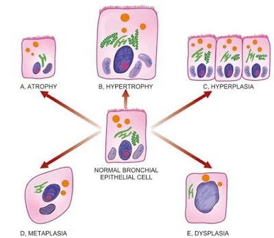
Definition :- Cell become change or adapt in response to adverse environmental changes that's called adaptation.

Types of adaptation
1. Atrophy (decrease cell size)
2. Dysplasia ( abnormal growth or development of cell )
3. Hyperplasia ( increase reproduction rate of the cell )
4. Hypertrophy ( increase cell size )
5. Metaplasia ( conversion of the cell from one type to another type e.g. squamus cell convert into columnar cell )

Adaptive Disorders

Difference between Metaplasia and Dysplasia


Similar Posts -


Dysplasia / Atypical hyperplasia

Definition – It is the disordered cellular development associated with metaplasia and hyperplasia caused by chronic irritation and prolonged inflammation. It is occur most commonly in epithelial cells.

Dysplasia characterised by cellular proliferation and cytologic changes as under…
  • Increase number of layers of epithelial cells.
  • Disorderly arrangement of cells from basal layer to the surface layer.
  • Loss of polarity i.e. nuclei lying away from basement membrane.
  • Cellular and nuclear pleomorphism.
  • Increase nucleocytoplasmic ratio.
  • Nuclear hyperchromatism.
  • Increase mitotic activity.

Most common examples of dysplastic changes are the uterine cervix and respiratory tract.

Metaplasia, dysplasia, adaptation , aasgaduli, pathology
Difference between Metaplasia and Dysplasia


Similar Posts -

Metaplasia


Definition – Metaplasia is a reversible condition in which one type of epithelial or mesenchymal adult cells convert into another type of epithelial or mesenchymal cells, usually in response to abnormal stimuli and it reverts back after removal of stimulus. If stimulus persists for long time then metaplasia convert into dysplasia and further convert into cancer.
Types – (1) Epithelial metaplasia (2) Mesenchymal metaplasia
1. Epithelial metaplasia – This is the more common type in which metaplastic changes may be patchy or diffuse and usually replaced by stronger but less well-specialised epithelium. There are two types of epithelial metaplasia…
A. SQUAMOUS METAPLASIA – It is caused by chronic irritation that may be mechanical, chemical or infective in origin. Some common examples…
  • In bronchus (normally lined by pseudostratified columnar ciliated epithelium) in chronic smokers.
  • In uterine endocervix (normally lined by simple columnar epithelium) in prolapsed of the uterus and in old age.
  • In gallbladder (normally lined by simple columnar epithelium) in chronic cholecystitis and cholelithiasis.
  • In prostate (ducts normally lined by simple columnar epithelium) in chronic prostatitis and oestrogen therapy.
  • In renal pelvis and urinary bladder (normally lined by transitional epithelium) in chronic infection and stones.
  • In vitamin A deficiency (xerophthalmia) in nose, bronchi, urinary tract, lacrimal and salivary glands.

B. COLUMNAR METAPLASIA – There are some conditions in which there is transformation to columnar epithelium. For example_
  • Intestinal metaplasia in healed chronic gastric ulcer.
  • Columnar metaplasia in Barrett’s oesophagus (change squamous to columnar epithelium).
  • Pseudostratified ciliated columnar epithelium change into columnar epithelium in chronic bronchitis and bronchiectasis.

2. Mesenchymal metaplasia – Less often, there is transformation of one adult type of mesenchymal tissue to another. There is two types
A. OSSEOUS METAPLASIA – Formation of bone in fibrous tissue, cartilage and myxoid tissue. Examples…
  • In arterial wall in old age (Monckeberg’s medial calcific sclerosis).
  • In soft tissues in myositis ossificans.
  • In cartilage of larynx and bronchi in elderly people.
  • In scar of chronic inflammation of prolonged duration.

B. CARTILAGINOUS METAPLASIA – It may occur in healing of fractures, where there is undue mobility.

Metaplasia, dysplasia, adaptation, pathology
Differences between Metaplasia and Dysplasia


Similar Posts -

Hyperplasia


Definition – Hyperplasia is an increase in the number of parenchymal cells resulting in enlargement of the organ or tissue. Quite often, both hyperplasia and hypertrophy occur together.
Causes – It may be physiological or pathological…
PHYSIOLOGICAL HYPERPLASIA –
                  1. Hormonal hyperplasia, it occurs due hormonal stimulation.
a.       Hyperplasia of female breast at puberty, during pregnancy and lactation.
b.      Hyperplasia of pregnant uterus.
c.       Proliferative activity of normal endometrium after a normal menstrual cycle.
d.      Prostatic hyperplasia in old age.
2. Compensatory hyperplasia i.e. it occurs due to removal of part of an organ or in the contralateral organ in paired organ e.g.
a.       Regeneration of liver after partial hepatectomy.
b.      Regeneration of epidermis after skin abrasion.
c.       Hyperplasia of nephrons after another nephrectomy.
PATHOLOGICAL HYPERPLASIA – It is occurs due to excessive stimulation of hormones or growth factors e.g.
  1. Endometrial hyperplasia due to excess oestrogen.
  2. In wound healing, there is formation of granulation tissue due to proliferation of fibroblasts and endothelial cells.
  3. Formation of skin warts from hyperplasia of epidermis due to human papilloma virus.
  4. Pseudocarcinomatous hyperplasia of the skin occurring at the margin of a non-healing ulcer.
  5. Intraductal epithelial hyperplasia in fibrocystic change in the breast.

MORPHOLOGICAL FEATURES –There is enlargement of the affected organ or tissue and increase in the number of cells due to increase DNA synthesis resulting in increase mitoses of the cells.
“Hyperplasia occurs in labile cells (epithelial cells of the skin, mucous membrane, lymph nodes, cells of bone marrow), stable cells (parenchymal cells of the liver, kidney, pancreas, adrenal, thyroid) while hyperplasia does not occurs in permanent cells (neurons, cardiac and skeletal muscles)”

cellular adaptation, pathology
Adaptive Disorders


Similar Posts -

Hypertrophy

Hypertrophy

Definition – Hypertrophy is an increase in the size of parenchymal cells resulting in enlargement of the organ or tissue, without any change in the number of cells.
Causes – It may be physiological or pathological…
PHYSIOLOGICAL HYPERTROPHY – Enlarged size of uterus in pregnancy.
PATHOLOGICAL HYPERTROPHY –
  1. Hypertrophy of cardiac muscles, e.g. systemic hypertension, aortic valve disease (stenosis and insufficiency), mitral insufficiency.
  2. Hypertrophy of smooth muscles, e.g. cardiac achalasia (in oesophagus), pyloric stenosis (in stomach), intestinal strictures, muscular arteries in hypertension.
  3. Hypertrophy of skeletal muscles, e.g. athletes and manual labourers.
  4. Compensatory hypertrophy after removal of contralateral organ, e.g. nephrectomy, adrenal hyperplasia.

Cardiac hypertrophy, adaptation, aasgaduli, pathology
Cardiac Hypertrophy


MORPHOLOGICAL FEATURES – The affected organ is enlarged and heavy. There is enlargement of muscles fibres as well as of nuclei. At ultra-structural level, there is increased synthesis of DNA and RNA, protein and increased number of organelles such as mitochondria, endoplasmic reticulum and myofibrils.


Similar Posts -

Infarction

Infarction

Definition – A necrotic area is called infarct and the process of infarct formation is called infarction.
Causes – Ischaemic necrosis (most common), stagnant hypoxia (venous obstruction), thrombosis, embolism, narrowing of the coronary artery (atherosclerosis) etc.
Types of infarcts –
1. According to colour …
            a. Pale or anemic infarct – It is due to arterial occlusion, commonly seen in compact organ like heart, kidney and spleen.
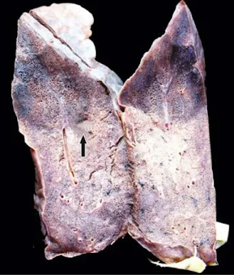
            b. Red or haemorrhagic infarct – It is due to obstruction of the artery, commonly seen in soft loose tissue e.g. lungs and intestine.

Infarct, haemorrhagic infarct, aasgaduli, pathology
Haemorrhagic Infarct of the Lung

2. According to the age … Recent and old.
3. According to the presence or absence of the infection…
            a. Bland – Free from bacterial contamination
            b. Infected – Infected with bacterial contamination.
4. According to the type of action…
            a. Bacteriostatic – It inhibit growth of bacteria e.g. tetracycline, erythromycin, sulfonamide etc.
            b. Bactericidal – It kills bacteria e.g. refampicin, penicillin etc.
5. According to the spectrum of activity… narrow and broad spectrum.
PATHOGENESIS –
  • Localized hyperaemia or congestion may occur due to obstruction of blood supply.
  • Within few hours edema and haemorrhage may occur.
  • Cellular change such as cloudy swelling appear early, necrosis occur within 12-15 hours.
  • Progressive proteolysis of necrotic tissue and there is lysis of red cells.
  • Acute inflammatory reactions appear in surrounding tissue.
  • Blood pigments like haematoidin and haemosiderin deposit in the infarct.
  • Following this granulation tissue starts to appears at the margin, finally infarct is replaced by collagen fibers and a scar is formed.

GROSSLY – Infarcts of solid arms are usually wedge shaped with apex at the obstructed artery and base of the surface of the organ. Most infarcts become pale due to breakdown of red cells but pulmonary infarcts never become pale due to extensive blood supply.

            Cerebral infarcts appear with central softening due to liquefaction necrosis (encephalomalacia), recent infarcts are generally slightly elevated over the surface but old infarcts are shrunken and depressed due to scar.

MICROSCOPICALLY – The tissue of affected area shows coagulation necrosis but in cerebral infarcts there is liquefaction necrosis. At the periphery of infarcts inflammatory reaction is noted. Finally the infarct is replaced by scar, but in cerebral infarcts there is gliosis.


Infarct, haemorrhagic infarct, aasgaduli, pathology
Infarcts of most commonly affected organ


Similar Posts -

Haemorrhage

Haemorrhage

Definition – Haemorrhage is the escape of blood from a blood vessel.
HAEMATOMA – Extravasation of blood into the tissue with resultant swelling is known as haematoma.
ECCHYMOSIS – Large extravasations of blood into the skin and mucous membranes are called ecchymosis.
PURPURA – Small areas of haemorrhage (up to 1 cm) into the skin and mucous membrane called purpura.
PETECHIAE – Minute pinhead size haemorrhages on the skin are called petechiae.
FORMS –
  1. Epistaxis  (nosebleed)
  2. Haemoptysis (coughing up blood)
  3. Haematemesis (vomiting blood)
  4. Haematuria (blood in urine)
  5. Melena (dark stool)
  6. Polymenorrhea (short menstrual cycle <21 days)
  7. Menorrhagia (heavy periods)
  8. Postpartum haemorrhage (excessive bleeding after childbirth)
  9. Antepartum haemorrhage (genital bleeding during pregnancy, third trimester to till birth or delivery)

Classification –
1. According to the nature or type … (a) External or visible haemorrhage (b) Internal or concealed haemorrhage.

2. According to the timing …
            a. Primary haemorrhage – When haemorrhage occurs just after injury of vessels.
            b. Reactionary haemorrhage – When bleeding within 24 hours (usually 4-6 hours) causing slipping of ligature, dislodgement of clot.
            c. Secondary haemorrhage – When haemorrhage occurs after 7-14 days causing surgery, sloughing of vessels due to infection.

3. According to the source…
            a. Arterial haemorrhage – When bleeding occurs due to rupture of artery. Blood is bright red in colour and emitted as spurting jet.
            b. Venous haemorrhage – When bleeding occurs due to rupture of veins. Blood is dark red in colour and emitted steady and copious flow.
            c. Capillary haemorrhage – When bleeding occurs due to rupture of capillaries. Blood is bright red in colour and emitted rapid oozing.

4. According to the duration … (a) Acute haemorrhage (b) Chronic haemorrhage

5. According to the type of intervention… (a) Surgical (b) Non-surgical haemorrhage

 Effects of Haemorrhage –
  • 33% of total blood, sudden loss may cause death.
  • 50% of total blood, loss over the period of 24 hours may not necessary fatal.
  • Chronic haemorrhage generally produce iron deficiency anaemia while occur acute haemorrhage may cause hypovolaemic shock.

Etiology –
  1. Trauma to the vessel wall, e.g. penetrating wound in the heart or great vessels.
  2. Spontaneous haemorrhage, e.g. rupture of an aneurism, septicaemia, bleeding diathesis, acute leukaemias, pernicious anaemia, scurvy.
  3. Inflammatory lesions of the vessel wall, e.g. bleeding from chronic peptic ulcer, typhoid ulcers, traversing a tuberculous cavity in the lung, syphilitic involvement of the aorta, polyarteritis nodosa.
  4. Neoplastic invasion, e.g. vascular invasion in carcinoma of the tongue.
  5. Vascular disease, e.g. atherosclerosis.
  6. Elevated pressure within the vessels, e.g. cerebral and retinal haemorrhage in systemic hypertension, severe haemorrhage from varicose veins due to high blood pressure in the veins of legs or oesophagus.

GHAI Essential Pediatrics

GHAI Essential Pediatrics (Eighth edition)


Books, GHAI, Pediatrics, Aasgaduli
GHAI Essential Pediatrics Book 8th edition


CONTENTS


1. Introduction to Pediatrics

2. Normal Growth and its Disorders – Somatic growth, physical growth, growth disorders, abnormalities of head size and shape.

Cunningham’s Mannual of practical anatomy vol-3 ( 16th edition )


2. Development – Normal development, behavioral disorders, habit disorders and tics.

4. Adolescent Health and Development – Physical aspect, cognitive and social development, problems faced by adolescents, role of health care provider.

5. Fluid and Electrolyte Disturbances – Composition of body fluids, deficit therapy, sodium, potassium, calcium, magnesium, acid-base disorders.


6. Nutrition – Macronutrients, normal diet, undernutrition, management of malnutrition.

7. Micronutrients in Health and Disease – Fat and water soluble vitamins, minerals and trace elements.

8. Newborn Infants – Resuscitation of a newborn, routine care, thermal protection, fluid and electrolyte management, kangaroo mother care, breastfeeding, care of low birth weight babies, infection in the neonates, perinatal asphyxia, respiratory distress, jaundice, congenital malformations, follow up of high risk neonates, metabolic disorders, effects of maternal condition on fetus and neonates.

         Park’s Textbook of PSM


9. Immunization and Immunodeficiency – Immunity, primary immunodeficiency disorders, immunization, commonly used vaccines, vaccine administration, immunization programs, immunization and special circumstances.

10. Infections and Infestations – Fever, common viral infections, viral hepatitis, HIV, common bacterial infections, tuberculosis, fungal infections, protozoal infections, congenital and perinatal infections, helminthic infestations.

Williams Gynecology


11. Disease of Gastrointestinal System and Liver – Gastrointestinal disorders, acute diarrhea, persistent diarrhoea, chronic diarrhoea, gastrointestinal bleeding, disorders of hepatobiliary system, acute viral hepatitis, liver failure, chronic liver disease.

12. Hematological Disorders – Anemia, approach to hemolytic anemia, hematopoietic stem cells transplantation, approach to a bleeding child, thrombotic disorders, leucocytosis, leucopenia.

13. Otolaryngology – Disease of the ear, nose, sinuses, oral cavity, pharynx, larynx, trachea, salivary glands.


14. Disorders of Respiratory System – Common respiratory symptoms, investigations for respiratory illness, respiratory tract infections, acute lower respiratory tract infections, bronchial asthma, foreign body aspiration, lung abscess, bronchiectasis, cystic fibrosis, ARDS.

15. Disorders of CVS – Congestive cardiac failure, congenital heart disease, acyanotic congenital heart defects, cyanotic heart disease, obstructive lesions, rheumatic fever and heart disease, infective endocarditis, myocardial disease, pericardial disease, systemic hypertension, pulmonary arterial hypertension, rhythm disorders, preventing adult cardiovascular disease.

Histology Brijesh Kumar


16. Disorders of Kidney and Urinary Tract – Renal anatomy and physiology, diagnostic evaluation, acute glomerulonephritis, nephrotic syndrome, chronic glomerulonephritis, urinary tract infections, acute kidney injury, chronic kidney disease, renal replacement therapy, disorders of renal tubular transport, enuresis, congenital abnormalities of kidney and urinary tract, cystic kidney disease.

17. Endocrine and Metabolic Disorders – Disorders of pituitary gland, thyroid gland, calcium metabolism, adrenal glands, obesity, disorders of gonadal hormones, diabetes mellitus.


18. Central Nervous System – Approach the neurological diagnosis, seizures, febrile convulsions, epilepsy, coma, acute bacterial meningitis, tuberculous meningitis, encephalitis and encephalopathies, intracranial space occupying lesions, subdural effusion, hydrocephalus, neural tube defects, acute hemiplegia of childhood, paraplegia and quadriplegia, ataxia, cerebral palsy, degenerative brain disorders, mental retardation, neurocutaneous syndromes.

19. Neuromuscular Disorders – Approach to evaluation, disorders affecting anterior horn cells, peripheral neuropathies, acute flaccid paralysis, neuromuscular junction disorders, muscle disorders.

Textbook of Pathology (Harsh Mohan) 7th edition

20. Childhood Malignancies – Leukemia, lymphoma, brain tumors, retinoblastoma, neuroblastoma, wilms tumors, malignant tumors of the liver, histiocytoses, oncologic emergencies, hematopoietic stem cells transplantation.

21. Rheumatological Disorders – Arthritis, systemic lupus erythematosus, juvenile dermatomyositis, scleroderma, mixed connective tissue disease, vasculitides.

22. Genetic Disorders – Chromosomal disorders, single gene disorders, polygenic inheritance, therapy of genetic disorders, prevention of genetic disorders.

Textbook of Pharmacology and Therapeutics


23. Inborn Errors of Metabolism – Suspecting an inborn error of metabolism, aminoacidopathies, urea cycle defects, organic acidurias, defects of carbohydrate metabolism, mitochondrial fatty acid oxidation defects, lysosomal storage disorders.

24. Eye Disorders – Pediatric eye screening, congenital and developmental abnormalities, acquired eye diseases.

25. Skin Disorders – Basic principles, genodermatoses, nevi, exczematous dermatitis, disorders of skin appendages, disorders of pigmentation, drug eruptions, infections, diseases caused by arthropods.

26. Poisoning, Injuries and Accidents – Poisoning, common poisoning, envenomation, injuries and accidents.

Physiology Sembulingam


27. Pediatric Critical Care – Assessment and monitoring of a seriously ill child, pediatric basic and advanced life support, shock, mechanical ventilation, nutrition in critically ill children, sedation, analgesia and paralysis, nosocomial infections in PICU, transfusions.

28. Common Medical Procedures – Obtaining blood specimens, removal of aspirated foreign body, nasogastric tube insertion, venous catheterization, capillary blood (hell prick), umbilical vessel catheterization, arterial catheterization, intraosseous infusion, lumbar puncture, thoracocentesis, abdominal paracentesis or ascetic tip, catheterization of bladder, peritoneal dialysis, bone marrow aspiration and biopsy, liver biopsy, renal biopsy.

Sabiston Textbook of Surgery


29. Rational Drug Therapy

30. Integrated Management of Neonatal and Childhood Illness

31. Rights of Children


ABOUT BOOK
Name Book
GHAI Essential Pediatrics 8th edition
Author/ Editior
Vinod K Paul, Arvind Bagga
__________Ramesh Agarwal, Naveen Sankhyan, Vandana Jain, Tushar R Godbole, Vijayalakshmi Bhatia, Kamran Afzal, Rakesh Lodha, Anuja Agarwal, Ashima Gulati, Ashok K Deorari, Aditi Sinha, Surjit Singh, Tanu Singhal, SK Kabra, Anshu Srivastava, Barath Jagadisan, SK Yachha, Tulika Seth, Sandeep Samant, Grant T Rohman, Jerome W Thompson, R Krishna Kumar, R Tandon, Manuj Raj,, PSN Menon, Anurag Bajpai, Kanika Ghai, Veena Kaira, Sheffali Gulati, Sadhna Shankar, Rachna Seth, Neerja Gupta, Madhulika Kabra, Neena Khanna, Seemab Rasool, P Ramesh Menon, Manjunatha Sarthi, Sidharth Kumar Sethi, Anu Thukral, AK Patwari, S Aneja, Rajeev Seth.
Type
pdf.
Size
67.60 MB
Pages
768
Pages type
Colored
Quality
Good

GHAI Essential Pediatrics Book Download from here

GHAI Essential Pediatiric Book Download from here

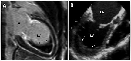
Endomyocardial Fibrosis